Masson's trichrome stain
Masson's Trichrome Stain is a histological staining technique used in the examination of tissue samples. It is named after the French pathologist Pierre Masson, who developed the technique in the early 20th century. This staining method is particularly useful for differentiating between collagen and muscle fibers, as well as for identifying increases in connective tissue. Masson's Trichrome Stain is widely used in the study of muscle diseases, fibrosis, and the liver.
Overview[edit]
Masson's Trichrome Stain involves the use of three different dyes: aniline blue, acid fuchsin, and Weigert's iron hematoxylin. The process results in nuclei being stained black by Weigert's iron hematoxylin, cytoplasm and muscle fibers in red/pink by acid fuchsin, and collagen in blue or green by aniline blue. This color differentiation allows for the detailed examination of tissue morphology and is particularly useful in identifying pathological changes.
Applications[edit]
The primary application of Masson's Trichrome Stain is in the field of pathology, where it is used to:
- Distinguish between muscle and collagen in tissue samples.
- Identify and assess the extent of fibrosis in various organs, particularly in the liver (e.g., cirrhosis) and kidney (e.g., glomerulonephritis).
- Study muscle diseases by highlighting the structural details of muscle tissue.
- Evaluate wound healing and the formation of scar tissue.
Procedure[edit]
The staining procedure for Masson's Trichrome involves several steps:
- Fixation of the tissue sample, typically in a solution like formalin.
- Embedding the tissue in paraffin and slicing it into thin sections.
- Deparaffinization and rehydration of tissue sections.
- Staining with Weigert's iron hematoxylin to color the nuclei.
- Staining with Biebrich scarlet-acid fuchsin solution to stain cytoplasm and muscle fibers.
- Differentiation in phosphomolybdic-phosphotungstic acid solution to remove the red stain except in muscle fibers.
- Staining with aniline blue to color the collagen.
- Dehydration, clearing, and mounting of the tissue sections for microscopic examination.
Advantages and Limitations[edit]
The advantages of Masson's Trichrome Stain include its ability to provide clear differentiation between collagen and muscle fibers, which is invaluable in diagnosing and studying various diseases. However, the technique has limitations, including the potential for variability in staining outcomes due to differences in the chemical composition of the dyes and the precise timing of the staining steps.
See Also[edit]
References[edit]
This medical article is a stub. You can help WikiMD by expanding the page. |
-
 Masson's trichrome stain of rat airway section
 -
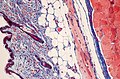 Masson's trichrome stain of skin tissue section
.jpg)


Medical Disclaimer: WikiMD is for informational purposes only and is not a substitute for professional medical advice. Content may be inaccurate or outdated and should not be used for diagnosis or treatment. Always consult your healthcare provider for medical decisions. Verify information with trusted sources such as CDC.gov and NIH.gov. By using this site, you agree that WikiMD is not liable for any outcomes related to its content. See full disclaimer.
Credits:Most images are courtesy of Wikimedia commons, and templates, categories Wikipedia, licensed under CC BY SA or similar.
Translate page: - East Asian
中文,
日本,
한국어,
South Asian
हिन्दी,
தமிழ்,
తెలుగు,
Urdu,
ಕನ್ನಡ,
Southeast Asian
Indonesian,
Vietnamese,
Thai,
မြန်မာဘာသာ,
বাংলা
European
español,
Deutsch,
français,
Greek,
português do Brasil,
polski,
română,
русский,
Nederlands,
norsk,
svenska,
suomi,
Italian
Middle Eastern & African
عربى,
Turkish,
Persian,
Hebrew,
Afrikaans,
isiZulu,
Kiswahili,
Other
Bulgarian,
Hungarian,
Czech,
Swedish,
മലയാളം,
मराठी,
ਪੰਜਾਬੀ,
ગુજરાતી,
Portuguese,
Ukrainian
