Pharyngeal groove
(Redirected from Sulcus pharyngei)
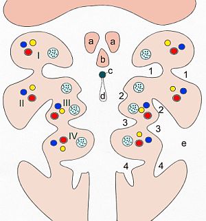
Pharyngeal grooves are anatomical structures found in the embryonic development of vertebrates. They are part of the pharyngeal arches, which are repetitive segments that appear on the lateral sides of the head and neck region of the embryo. The pharyngeal grooves, also known as branchial grooves or clefts, are external indentations located between the pharyngeal arches. In fish and some amphibians, these grooves develop into gills, which are essential for breathing underwater. However, in mammals, including humans, the pharyngeal grooves do not develop into gills but instead contribute to the formation of various structures in the head and neck.
Development and Significance[edit | edit source]
During embryonic development, the pharyngeal grooves form on the outer surface of the embryo, while the pharyngeal pouches develop on the inner surface, facing the pharynx. In humans, there are typically four pairs of pharyngeal grooves. The first pharyngeal groove plays a significant role in the development of the ear, contributing to the formation of the external auditory meatus and the external part of the tympanic membrane. The remaining grooves usually regress and do not form distinct adult structures, but they can contribute to the development of certain neck tissues and structures.
Clinical Significance[edit | edit source]
In some cases, remnants of the pharyngeal grooves may persist, leading to congenital anomalies such as branchial cleft cysts or fistulas. These conditions result from the incomplete obliteration of the pharyngeal grooves and can lead to the formation of cysts or abnormal passages that can become infected. Surgical removal is often required to treat these anomalies.
Comparison with Pharyngeal Pouches[edit | edit source]
While the pharyngeal grooves are located externally, the pharyngeal pouches are their internal counterparts. The pouches also play crucial roles in the development of structures within the head and neck. For example, the first pharyngeal pouch contributes to the formation of the Eustachian tube, middle ear cavity, and the inner layer of the tympanic membrane. The interactions between the pharyngeal grooves and pouches are essential for the proper development of the pharyngeal arch derivatives.
Evolutionary Perspective[edit | edit source]
The presence of pharyngeal grooves is a testament to the shared evolutionary history among vertebrates. In aquatic organisms like fish, these structures evolve into functional gills, highlighting their role in respiration. In terrestrial vertebrates, the transformation of these embryonic structures reflects the adaptation to an air-breathing environment, showcasing the versatility and evolutionary significance of the pharyngeal arch system.
Search WikiMD
Ad.Tired of being Overweight? Try W8MD's physician weight loss program.
Semaglutide (Ozempic / Wegovy and Tirzepatide (Mounjaro / Zepbound) available.
Advertise on WikiMD
|
WikiMD's Wellness Encyclopedia |
| Let Food Be Thy Medicine Medicine Thy Food - Hippocrates |
Translate this page: - East Asian
中文,
日本,
한국어,
South Asian
हिन्दी,
தமிழ்,
తెలుగు,
Urdu,
ಕನ್ನಡ,
Southeast Asian
Indonesian,
Vietnamese,
Thai,
မြန်မာဘာသာ,
বাংলা
European
español,
Deutsch,
français,
Greek,
português do Brasil,
polski,
română,
русский,
Nederlands,
norsk,
svenska,
suomi,
Italian
Middle Eastern & African
عربى,
Turkish,
Persian,
Hebrew,
Afrikaans,
isiZulu,
Kiswahili,
Other
Bulgarian,
Hungarian,
Czech,
Swedish,
മലയാളം,
मराठी,
ਪੰਜਾਬੀ,
ગુજરાતી,
Portuguese,
Ukrainian
Medical Disclaimer: WikiMD is not a substitute for professional medical advice. The information on WikiMD is provided as an information resource only, may be incorrect, outdated or misleading, and is not to be used or relied on for any diagnostic or treatment purposes. Please consult your health care provider before making any healthcare decisions or for guidance about a specific medical condition. WikiMD expressly disclaims responsibility, and shall have no liability, for any damages, loss, injury, or liability whatsoever suffered as a result of your reliance on the information contained in this site. By visiting this site you agree to the foregoing terms and conditions, which may from time to time be changed or supplemented by WikiMD. If you do not agree to the foregoing terms and conditions, you should not enter or use this site. See full disclaimer.
Credits:Most images are courtesy of Wikimedia commons, and templates, categories Wikipedia, licensed under CC BY SA or similar.
Contributors: Prab R. Tumpati, MD

