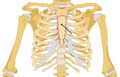
The Sternum
Anatomy > Gray's Anatomy of the Human Body > II. Osteology > The Strenum.
4a. The Sternum[edit]
(Breast Bone)

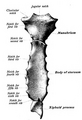
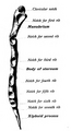
The sternum (Figs. 115 to 117) is an elongated, flattened bone, forming the middle portion of the anterior wall of the thorax. Its upper end supports the clavicles, and its margins articulate with the cartilages of the first seven pairs of ribs. It consists of three parts, named from above downward, the manubrium, the body or gladiolus, and the xiphoid process; in early life the body consists of four segments or sternebroe In its natural position the inclination of the bone is oblique from above, downward and forward.
It is slightly convex in front and concave behind; broad above, becoming narrowed at the point where the manubrium joins the body, after which it again widens a little to below the middle of the body, and then narrows to its lower extremity. Its average length in the adult is about 17 cm., and is rather greater in the male than in the female.
Manubrium (manubrium sterni)[edit]
The manubrium is of a somewhat quadrangular form, broad and thick above, narrow below at its junction with the body.
Surfaces[edit]
Its anterior surface convex from side to side, concave from above downward, is smooth, and affords attachment on either side to the sternal origins of the Pectoralis major and Sternocleidomastoideus. Sometimes the ridges limiting the attachments of these muscles are very distinct. Its posterior surface concave and smooth, affords attachment on either side to the Sternohyoideus and Sternothyreoideus.


Borders[edit]
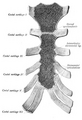
The superior border is the thickest and presents at its center the jugular or presternal notch on either side of the notch is an oval articular surface, directed upward, backward, and lateralward, for articulation with the sternal end of the clavicle. The inferior border oval and rough, is covered in a fresh state with a thin layer of cartilage, for articulation with the body.
The lateral borders are each marked above by a depression for the first costal cartilage, and below by a small facet, which, with a similar facet on the upper angle of the body, forms a notch for the reception of the costal cartilage of the second rib. Between the depression for the first costal cartilage and the demi-facet for the second is a narrow, curved edge, which slopes from above downward and medialward.
Body (corpus sterni; gladiolus)[edit]
The body, considerably longer, narrower, and thinner than the manubrium, attains its greatest breadth close to the lower end.
Surfaces[edit]
Its anterior surface is nearly flat, directed upward and forward, and marked by three transverse ridges which cross the bone opposite the third, fourth, and fifth articular depressions. 18 It affords attachment on either side to the sternal origin of the Pectoralis major. At the junction of the third and fourth pieces of the body is occasionally seen an orifice, the sternal foramen of varying size and form.
The posterior surface slightly concave, is also marked by three transverse lines, less distinct, however, than those in front; from its lower part, on either side, the Transversus thoracis takes origin.
Borders[edit]
The superior border is oval and articulates with the manubrium, the junction of the two forming the sternal angle (angulus Ludovici 19). The inferior border is narrow, and articulates with the xiphoid process. Each lateral border (Fig. 117), at its superior angle, has a small facet, which with a similar facet on the manubrium, forms a cavity for the cartilage of the second rib; below this are four angular depressions which receive the cartilages of the third, fourth, fifth, and sixth ribs, while the inferior angle has a small facet, which, with a corresponding one on the xiphoid process, forms a notch for the cartilage of the seventh rib.
These articular depressions are separated by a series of curved interarticular intervals, which diminish in length from above downward, and correspond to the intercostal spaces. Most of the cartilages belonging to the true ribs, as will be seen from the foregoing description, articulate with the sternum at the lines of junction of its primitive component segments. This is well seen in many of the lower animals, where the parts of the bone remain ununited longer than in man.
Xiphoid Process[edit]
(processus xiphoideus; ensiform or xiphoid appendix)
The xiphoid process is the smallest of the three pieces: it is thin and elongated, cartilaginous in structure in youth, but more or less ossified at its upper part in the adult.
Surfaces[edit]
Its anterior surface affords attachment on either side to the anterior costoxiphoid ligament and a small part of the Rectus abdominis; its posterior surface to the posterior costoxiphoid ligament and to some of the fibers of the diaphragm and Transversus thoracis, its lateral borders to the aponeuroses of the abdominal muscles.
Above, it articulates with the lower end of the body, and on the front of each superior angle presents a facet for part of the cartilage of the seventh rib; below, by its pointed extremity, it gives attachment to the linea alba. The xiphoid process varies much in form; it may be broad and thin, pointed, bifid, perforated, curved, or deflected considerably to one or other side.
Structure[edit]
The sternum is composed of highly vascular cancellous tissue, covered by a thin layer of compact bone which is thickest in the manubrium between the articular facets for the clavicles.
Ossification[edit]
The sternum originally consists of two cartilaginous bars, situated one on either side of the median plane and connected with the cartilages of the upper nine ribs of its own side. These two bars fuse with each other along the middle line to form the cartilaginous sternum which is ossified from six centers: one for the manubrium, four for the body, and one for the xiphoid process (Fig. 118).
The ossific centers appear in the intervals between the articular depressions for the costal cartilages, in the following order: in the manubrium and first piece of the body, during the sixth month; in the second and third pieces of the body, during the seventh month of fetal life; in its fourth piece, during the first year after birth; and in the xiphoid process, between the fifth and eighteenth years.
The centers make their appearance at the upper parts of the segments, and proceed gradually downward. 20 To these may be added the occasional existence of two small episternal centers, which make their appearance one on either side of the jugular notch; they are probably vestiges of the episternal bone of the monotremata and lizards.
Occasionally some of the segments are formed from more than one center, the number and position of which vary (Fig. 120). Thus, the first piece may have two, three, or even six centers. When two are present, they are generally situated one above the other, the upper being the larger; the second piece has seldom more than one; the third, fourth, and fifth pieces are often formed from two centers placed laterally, the irregular union of which explains the rare occurrence of the sternal foramen (Fig. 121), or of the vertical fissure which occasionally intersects this part of the bone constituting the malformation known as fissura sterni these conditions are further explained by the manner in which the cartilaginous sternum is formed.
More rarely still the upper end of the sternum may be divided by a fissure. Union of the various centers of the body begins about puberty, and proceeds from below upward (Fig. 119); by the age of twenty-five they are all united. The xiphoid process may become joined to the body before the age of thirty, but this occurs more frequently after forty; on the other hand, it sometimes remains ununited in old age.
In advanced life the manubrium is occasionally joined to the body by bone. When this takes place, however, the bony tissue is generally only superficial, the central portion of the intervening cartilage remaining unossified.


Articulations[edit]
The sternum articulates on either side with the clavicle and upper seven costal cartilages. Note 18 Paterson (The Human Sternum, 1904), who examined 524 specimens, points out that these ridges are altogether absent in 26.7 per cent.; that in 69 per cent. a ridge exists opposite the third costal attachment; in 39 per cent.
opposite the fourth; and in 4 per cent. only, opposite the fifth. Note 19 Named after the French surgeon Antoine Louis, 1723–1792. The Latin name angulus Ludovici is not infrequently mistranslated into English as “the angle of Ludwig.” Note 20 Out of 141 sterna between the time of birth and the age of sixteen years, Paterson (op. cit) found the fourth or lowest center for the body present only in thirty-eight cases—i. e 26.9 per cent.
Additional Images[edit]
-
Position of sternum (shown in red). Animation.
-
 Sternum seen posteriorly.
 -
 Sternum cut along the frontal plane showing interior of the bone.
 -
 Sternum, lateral aspect.
 -
 Position of the sternum the thoracic cage.


Gray's Anatomy[edit]
- Gray's Anatomy Contents
- Gray's Anatomy Subject Index
- About Classic Gray's Anatomy
- Glossary of anatomy terms
Anatomy atlases (external)[edit]
[1] - Anatomy Atlases
| Human systems and organs | ||||||||||||||
|---|---|---|---|---|---|---|---|---|---|---|---|---|---|---|
|

Medical Disclaimer: WikiMD is for informational purposes only and is not a substitute for professional medical advice. Content may be inaccurate or outdated and should not be used for diagnosis or treatment. Always consult your healthcare provider for medical decisions. Verify information with trusted sources such as CDC.gov and NIH.gov. By using this site, you agree that WikiMD is not liable for any outcomes related to its content. See full disclaimer.
Credits:Most images are courtesy of Wikimedia commons, and templates, categories Wikipedia, licensed under CC BY SA or similar.
Translate page: - East Asian
中文,
日本,
한국어,
South Asian
हिन्दी,
தமிழ்,
తెలుగు,
Urdu,
ಕನ್ನಡ,
Southeast Asian
Indonesian,
Vietnamese,
Thai,
မြန်မာဘာသာ,
বাংলা
European
español,
Deutsch,
français,
Greek,
português do Brasil,
polski,
română,
русский,
Nederlands,
norsk,
svenska,
suomi,
Italian
Middle Eastern & African
عربى,
Turkish,
Persian,
Hebrew,
Afrikaans,
isiZulu,
Kiswahili,
Other
Bulgarian,
Hungarian,
Czech,
Swedish,
മലയാളം,
मराठी,
ਪੰਜਾਬੀ,
ગુજરાતી,
Portuguese,
Ukrainian
