Coarctation of the aorta
Editor-In-Chief: Prab R Tumpati, MD
Obesity, Sleep & Internal medicine
Founder, WikiMD Wellnesspedia &
W8MD medical weight loss NYC and sleep center NYC
| Coarctation of the aorta | |
|---|---|

| |
| Synonyms | Aortic coarctation |
| Pronounce | N/A |
| Specialty | N/A |
| Symptoms | High blood pressure, headache, muscle weakness, leg cramps, cold feet |
| Complications | Heart failure, aortic aneurysm, stroke, coronary artery disease |
| Onset | Childhood or adulthood |
| Duration | Chronic |
| Types | N/A |
| Causes | Congenital heart defect |
| Risks | Turner syndrome, bicuspid aortic valve, family history |
| Diagnosis | Echocardiogram, chest X-ray, MRI, CT scan |
| Differential diagnosis | Hypertension, aortic stenosis, interrupted aortic arch |
| Prevention | N/A |
| Treatment | Surgery, balloon angioplasty, stenting |
| Medication | Antihypertensive drugs |
| Prognosis | Generally good with treatment |
| Frequency | 4 in 10,000 newborns |
| Deaths | Rare with treatment |
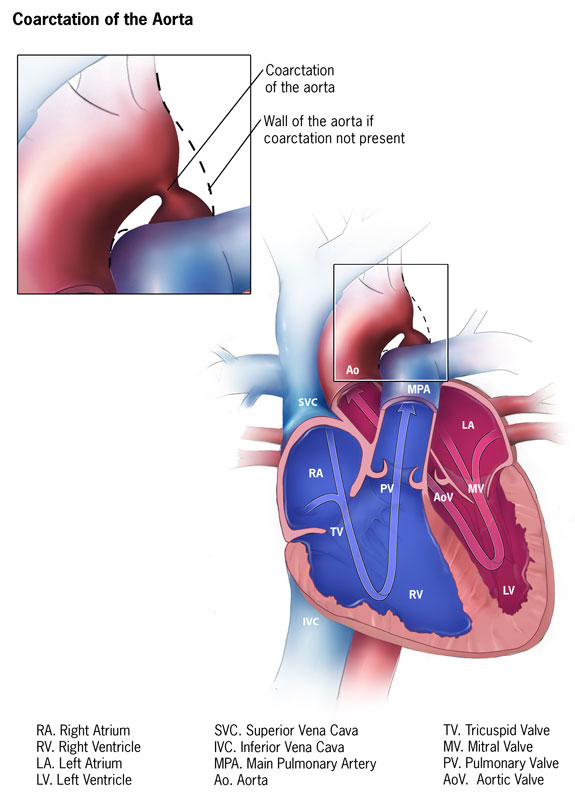
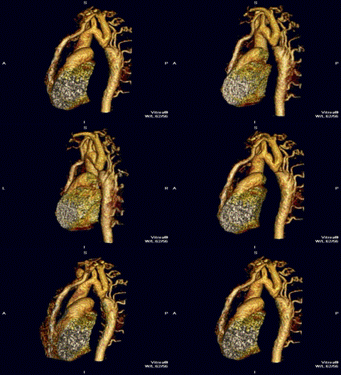
Coarctation of the aorta is a birth defect where part of the aorta, is narrower than usual.
Pronunciation[edit | edit source]
Coarctation of the aorta is pronounced koh-ark-TEY-shun
Birth defect[edit | edit source]
Coarctation of the aorta is a birth defect in which a part of the aorta is narrower than usual.
Needs surgery[edit | edit source]
If the narrowing is severe enough and if it is not diagnosed, the baby may have serious problems and may need surgery or other procedures soon after birth. For this reason, coarctation of the aorta is often considered a critical congenital heart defect.
Cause[edit | edit source]
The defect occurs when a baby’s aorta does not form correctly as the baby grows and develops during pregnancy. The exact cause is not known.
Location[edit | edit source]
The narrowing of the aorta usually happens in the part of the blood vessel just after the arteries branch off to take blood to the head and arms, near the patent ductus arteriosus, although sometimes the narrowing occurs before or after the ductus arteriosus.
Ductus arteriosus[edit | edit source]
- In some babies with coarctation, it is thought that some tissue from the wall of ductus arteriosus blends into the tissue of the aorta.
- When the tissue tightens and allows the ductus arteriosus to close normally after birth, this extra tissue may also tighten and narrow the aorta.
Mechanism[edit | edit source]
The narrowing, or coarctation, blocks normal blood flow to the body. This can back up flow into the left ventricle of the heart, making the muscles in this ventricle work harder to get blood out of the heart.
Effects on blood flow[edit | edit source]
Since the narrowing of the aorta is usually located after arteries branch to the upper body, coarctation in this region can lead to normal or high blood pressure and pulsing of blood in the head and arms and low blood pressure and weak pulses in the legs and lower body. If the condition is very severe, enough blood may not be able to get through to the lower body. The extra work on the heart can cause the walls of the heart to become thicker in order to pump harder. This eventually weakens the heart muscle. If the aorta is not widened, the heart may weaken enough that it leads to heart failure.
Other congenital heart defects[edit | edit source]
Coarctation of the aorta often occurs with other congenital heart defects.
Incidence[edit | edit source]
- In a 2019 study, using 2010-2014 data from 39 population-based birth defects surveillance programs, the Centers for Disease Control and Prevention (CDC) estimated that about 4 of every 10,000 babies born had coarctation of the aorta.
- This means about 2,200 babies are born each year in the United States with coarctation of the aorta.
- In other words, about 1 in every 1,800 babies born in the United States each year are born with coarctation of the aorta.
Risk Factors[edit | edit source]
While the exact cause for coarctation of the aorta is unknown, some babies have heart defects because of changes in their genes or chromosomes.
Role of genes and environment[edit | edit source]
Heart defects, like coarctation of the aorta, are also thought to be caused by a combination of genes and other risk factors, such as things the mother comes in contact with in the environment, what the mother eats or drinks, or medicines the mother uses.
Diagnosis[edit | edit source]
Coarctation of the aorta is usually diagnosed after the baby is born. How early in life the defect is diagnosed usually depends on how mild or severe the symptoms are.
Newborn screening[edit | edit source]
Newborn screening using pulse oximetry during the first few days of life may or may not detect coarctation of the aorta. In babies with a more serious condition, early signs usually include:
- pale skin
- irritability
- heavy sweating
- difficulty breathing
Physical exam[edit | edit source]
- Detection of the defect is often made during a physical exam.
- In infants and older individuals, the pulse will be noticeably weaker in the legs or groin than it is in the arms or neck, and a heart murmur—an abnormal whooshing sound caused by disrupted blood flow—may be heard through a doctor’s stethoscope.
- Older children and adults with coarctation of the aorta often have high blood pressure in the arms.
Tests - echocardiogram[edit | edit source]
- Once suspected, an echocardiogram is the most commonly used test to confirm the diagnosis.
- An echocardiogram will show the location and severity of the coarctation and whether any other heart defects are present.
Other tests[edit | edit source]
Other tests to measure the function of the heart may be used including chest x-ray, electrocardiogram (EKG), magnetic resonance imaging (MRI), and cardiac catheterization.
Critical CHD[edit | edit source]
- Coarctation of the aorta is often considered a critical congenital heart defect (critical CHD) because if the narrowing is severe enough and it is not diagnosed, the baby may have serious problems soon after birth.
- CCHDs also can be detected with newborn pulse oximetry screening.
Treatments[edit | edit source]
No matter what age the defect is diagnosed, the narrow aorta will need to be widened surgically once symptoms are present. This can be done with surgery or a procedure called balloon angioplasty, which is done during a cardiac catheterization.
Balloon angioplasty[edit | edit source]
A balloon angioplasty is a procedure that uses a thin, flexible tube, called a catheter, which is inserted into a blood vessel and directed to the aorta. When the catheter reaches the narrow area of the aorta, a balloon at the tip is inflated to expand the blood vessel.
Stents[edit | edit source]
- Sometimes a mesh-covered tube (stent) is inserted to keep the vessel open.
- The stent is used more often to initially widen the aorta or re-widen it if the aorta narrows again after surgery has been performed.
Surgical reconstruction of aorta[edit | edit source]
During surgery to correct a coarctation, the narrow portion is removed and the aorta is reconstructed or patched to allow blood to flow normally through the aorta.
Monitor BP[edit | edit source]
- Even after surgery, children with a coarctation of the aorta often have high blood pressure that is treated with medicine.
- It is important for children and adults with coarctation of the aorta to follow up regularly with a cardiologist to monitor their progress and check for other health conditions that might develop as they get older.
Latest articles - Coarctation of the aorta
Frequently asked question on Coarctation of the aorta[edit source]
- Have question on Coarctation of the aorta? Ask in the Question portal.
- FAQ's on Coarctation of the aorta
- What is a characteristic sign of coarctation of the aorta?
High blood pressure especially in the upper body, low blood pressure or weak pulses in the lower part of the body, muscle weakness, headaches, etc.
- How do you assess for coarctation of the aorta?
In addition to a physical examination, tests such as echocardiogram, CT chest, X-rays, and other tests can help.
- What causes coarctation of the aorta?
The exact cause is not known. It is likely a combination of genes and other risk factors, such as things the mother comes in contact with in the environment, what the mother eats or drinks, or medicines the mother uses
- How rare is coarctation of the aorta?
1 in every 1,800 babies born in the United States each year are born with coarctation of the aorta.
- What can happen if the coarctation is not repaired?
It can lead to heart failure
- How long does surgery for coarctation of the aorta take?
While it depends, typically, it takes about 2-3 hours.
Answer these questions[edit | edit source]
- What is an expected assessment finding in a child with coarctation of the aorta?
- Where is coarctation of the aorta COA located?
- Can coarctation be detected before birth?
- Which artery is enlarged in coarctation of aorta?
- How does a PDA help coarctation of the aorta?
- Why does aortic coarctation cause rib notching?
- What does narrowing of the aorta mean?
Coarctation of the aorta on Wikipedia[edit source]
| Congenital vascular defects / Vascular malformation | ||||||
|---|---|---|---|---|---|---|
|
Search WikiMD
Ad.Tired of being Overweight? Try W8MD's physician weight loss program.
Semaglutide (Ozempic / Wegovy and Tirzepatide (Mounjaro / Zepbound) available.
Advertise on WikiMD
|
WikiMD's Wellness Encyclopedia |
| Let Food Be Thy Medicine Medicine Thy Food - Hippocrates |
Translate this page: - East Asian
中文,
日本,
한국어,
South Asian
हिन्दी,
தமிழ்,
తెలుగు,
Urdu,
ಕನ್ನಡ,
Southeast Asian
Indonesian,
Vietnamese,
Thai,
မြန်မာဘာသာ,
বাংলা
European
español,
Deutsch,
français,
Greek,
português do Brasil,
polski,
română,
русский,
Nederlands,
norsk,
svenska,
suomi,
Italian
Middle Eastern & African
عربى,
Turkish,
Persian,
Hebrew,
Afrikaans,
isiZulu,
Kiswahili,
Other
Bulgarian,
Hungarian,
Czech,
Swedish,
മലയാളം,
मराठी,
ਪੰਜਾਬੀ,
ગુજરાતી,
Portuguese,
Ukrainian
Medical Disclaimer: WikiMD is not a substitute for professional medical advice. The information on WikiMD is provided as an information resource only, may be incorrect, outdated or misleading, and is not to be used or relied on for any diagnostic or treatment purposes. Please consult your health care provider before making any healthcare decisions or for guidance about a specific medical condition. WikiMD expressly disclaims responsibility, and shall have no liability, for any damages, loss, injury, or liability whatsoever suffered as a result of your reliance on the information contained in this site. By visiting this site you agree to the foregoing terms and conditions, which may from time to time be changed or supplemented by WikiMD. If you do not agree to the foregoing terms and conditions, you should not enter or use this site. See full disclaimer.
Credits:Most images are courtesy of Wikimedia commons, and templates, categories Wikipedia, licensed under CC BY SA or similar.
Contributors: Prab R. Tumpati, MD


