Computer-aided diagnosis
Computer-aided diagnosis (CAD), also known as computer-aided detection, is a procedure in the field of medicine that uses computer algorithms to assist doctors in the interpretation of medical images. Imaging techniques in radiology, magnetic resonance imaging (MRI), ultrasound, and computed tomography (CT) can benefit from CAD systems in identifying diseases such as cancer (e.g., breast cancer, lung cancer), by highlighting conspicuous sections, thus drawing the attention of the radiologist to these areas.
History[edit]
The development of CAD systems began in the early 1960s with the advent of digital computing. The first applications were focused on assisting radiologists in detecting breast cancer on mammograms. Over the years, the technology has evolved significantly, incorporating more sophisticated machine learning and artificial intelligence algorithms to improve detection rates and reduce false positives.
How CAD Works[edit]
CAD systems process digital images from various medical imaging techniques. The process involves several steps: 1. Image Acquisition: The digital medical images are acquired from the imaging equipment. 2. Image Pre-processing: These images are then enhanced for better visualization, which may involve noise reduction and contrast enhancement. 3. Feature Extraction: The system identifies and isolates specific features within the images that are characteristic of particular diseases. 4. Analysis and Pattern Recognition: Using machine learning algorithms, the system analyzes these features to detect anomalies. 5. Marking: The suspected areas are marked on the images, providing visual cues for the radiologist to examine further.
Applications[edit]
CAD systems are used in various fields of medical imaging. Some of the notable applications include: - Mammography: CAD systems are widely used to detect early signs of breast cancer, such as microcalcifications and masses. - Lung Cancer: In chest radiography and CT scans, CAD systems help in identifying nodules that may indicate lung cancer. - Colon Cancer: In virtual colonoscopy, CAD can assist in detecting polyps. - Liver Cancer: CAD systems are used in CT and MRI scans to identify liver lesions. - Brain Tumors: MRI-based CAD systems assist in the detection and delineation of brain tumors.
Benefits and Limitations[edit]
- Benefits
- Increased Detection Rate: CAD can help in identifying lesions that may be overlooked by the human eye. - Consistency: CAD systems provide a consistent second opinion to radiologists. - Efficiency: They can reduce the time required for image analysis, aiding in faster diagnosis.
- Limitations
- False Positives: One of the major challenges of CAD systems is the high rate of false positives, which can lead to unnecessary biopsies and anxiety. - Dependence: There is a risk of over-reliance on CAD systems by radiologists, potentially undermining their diagnostic skills. - Cost: The high cost of CAD systems can be a barrier to their widespread adoption.
Future Directions[edit]
The future of CAD lies in the integration of more advanced AI and deep learning technologies, which promise to improve the accuracy and reduce the false positive rates of these systems. Additionally, the development of CAD systems for new applications and imaging modalities continues to be an active area of research.
See Also[edit]
This medical article is a stub. You can help WikiMD by expanding the page. |
-
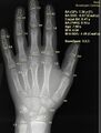 X-ray of hand with bone age analysis by BoneXpert software
 -
IBM Medical Sieve
-
Histogram equalization
-
Support vector machine


Medical Disclaimer: WikiMD is for informational purposes only and is not a substitute for professional medical advice. Content may be inaccurate or outdated and should not be used for diagnosis or treatment. Always consult your healthcare provider for medical decisions. Verify information with trusted sources such as CDC.gov and NIH.gov. By using this site, you agree that WikiMD is not liable for any outcomes related to its content. See full disclaimer.
Credits:Most images are courtesy of Wikimedia commons, and templates, categories Wikipedia, licensed under CC BY SA or similar.
Translate page: - East Asian
中文,
日本,
한국어,
South Asian
हिन्दी,
தமிழ்,
తెలుగు,
Urdu,
ಕನ್ನಡ,
Southeast Asian
Indonesian,
Vietnamese,
Thai,
မြန်မာဘာသာ,
বাংলা
European
español,
Deutsch,
français,
Greek,
português do Brasil,
polski,
română,
русский,
Nederlands,
norsk,
svenska,
suomi,
Italian
Middle Eastern & African
عربى,
Turkish,
Persian,
Hebrew,
Afrikaans,
isiZulu,
Kiswahili,
Other
Bulgarian,
Hungarian,
Czech,
Swedish,
മലയാളം,
मराठी,
ਪੰਜਾਬੀ,
ગુજરાતી,
Portuguese,
Ukrainian
