The Muscles of the Mouth
Anatomy > Gray's Anatomy of the Human Body > IV. Myology > 4d. The Muscles of the Mouth
Henry Gray (1821–1865). Anatomy of the Human Body. 1918.
The Muscles of the Mouth[edit | edit source]
The muscles of the mouth are:
- Quadratus labii superioris
- Quadratus labii inferioris
- Caninus
- Triangularis
- Zygomaticus
- Buccinator
- Mentalis
- Orbicularis oris
- Risorius
Quadratus labii superioris[edit | edit source]
The Quadratus labii superioris is a broad sheet, the origin of which extends from the side of the nose to the zygomatic bone. Its medial fibers form the angular head which arises by a pointed extremity from the upper part of the frontal process of the maxilla and passing obliquely downward and lateralward divides into two slips. One of these is inserted into the greater alar cartilage and skin of the nose; the other is prolonged into the lateral part of the upper lip, blending with the infraorbital head and with the Orbicularis oris.
The intermediate portion or infraorbital head arises from the lower margin of the orbit immediately above the infraorbital foramen, some of its fibers being attached to the maxilla, others to the zygomatic bone. Its fibers converge, to be inserted into the muscular substance of the upper lip between the angular head and the Caninus. The lateral fibers, forming the zygomatic head arise from the malar surface of the zygomatic bone immediately behind the zygomaticomaxillary suture and pass downward and medialward to the upper lip.
Caninus[edit | edit source]
The Caninus (Levator anguli oris) arises from the canine fossa, immediately below the infraorbital foramen; its fibers are inserted into the angle of the mouth, intermingling with those of the Zygomaticus, Triangularis, and Orbicularis oris.
Zygomaticus[edit | edit source]
The Zygomaticus (Zygomaticus major) arises from the zygomatic bone, in front of the zygomaticotemporal suture, and descending obliquely with a medial inclination, is inserted into the angle of the mouth, where it blends with the fibers of the Caninus, Orbicularis oris, and Triangularis.
Nerves
This group of muscles is supplied by the facial nerve.
ActionsThe Quadratus labii superioris is the proper elevator of the upper lip, carrying it at the same time a little forward. Its angular head acts as a dilator of the naris; the infraorbital and zygomatic heads assist in forming the nasolabial furrow, which passes from the side of the nose to the upper lip and gives to the face an expression of sadness. When the whole muscle is in action it gives to the countenance an expression of contempt and disdain. The Quadratus labii superioris raises the angle of the mouth and assists the Caninus in producing the nasolabial furrow. The Zygomaticus draws the angle of the mouth backward and upward, as in laughing.
Mentalis[edit | edit source]
The Mentalis (Levator menti) is a small conical fasciculus, situated at the side of the frenulum of the lower lip. It arises from the incisive fossa of the mandible, and descends to be inserted into the integument of the chin.
Quadratus labii inferioris[edit | edit source]
The Quadratus labii inferioris (Depressor labii inferioris; Quadratus menti) is a small quadrilateral muscle. It arises from the oblique line of the mandible, between the symphysis and the mental foramen, and passes upward and medialward, to be inserted into the integument of the lower lip, its fibers blending with the Orbicularis oris, and with those of its fellow of the opposite side. At its origin it is continuous with the fibers of the Platysma. Much yellow fat is intermingled with the fibers of this muscle.
Triangularis[edit | edit source]
The Triangularis (Depressor anguli oris) arises from the oblique line of the mandible, whence its fibers converge, to be inserted by a narrow fasciculus, into the angle of the mouth. At its origin it is continuous with the Platysma, and at its insertion with the Orbicularis oris and Risorius; some of its fibers are directly continuous with those of the Caninus, and others are occasionally found crossing from the muscle of one side to that of the other; these latter fibers constitute the Transversus menti
Nerves - This group of muscles is supplied by the facial nerve.
Actions The Mentalis raises and protrudes the lower lip, and at the same time wrinkles the skin of the chin, expressing doubt or disdain. The Quadratus labii inferioris draws the lower lip directly downward and a little lateralward, as in the expression of irony. The Triangularis depresses the angle of the mouth, being the antagonist of the Caninus and Zygomaticus; acting with the Caninus, it will draw the angle of the mouth medialward. The Platysma which retracts and depresses the angle of the mouth belongs with this group.
Buccinator[edit | edit source]
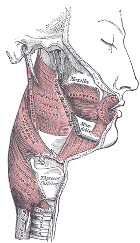
The Buccinator (Fig. 380) is a thin quadrilateral muscle, occupying the interval between the maxilla and the mandible at the side of the face. It arises from the outer surfaces of the alveolar processes of the maxilla and mandible, corresponding to the three molar teeth; and behind, from the anterior border of the pterygomandibular raphé which separates it from the Constrictor pharyngis superior. The fibers converge toward the angle of the mouth, where the central fibers intersect each other, those from below being continuous with the upper segment of the Orbicularis oris, and those from above with the lower segment; the upper and lower fibers are continued forward into the corresponding lip without decussation.
Relations The Buccinator is covered by the buccopharyngeal fascia, and is in relation by its superficial surface behind, with a large mass of fat, which separates it from the ramus of the mandible, the Masseter, and a small portion of the Temporalis; this fat has been named the suctorial pad because it is supposed to assist in the act of sucking. The parotid duct pierces the Buccinator opposite the second molar tooth of the maxilla. The deep surface is in relation with the buccal glands and mucous membrane of the mouth.
pterygomandibular raphé[edit | edit source]
The (pterygomandibular ligament) is a tendinous band of the buccopharyngeal fascia, attached by one extremity to the hamulus of the medial pterygoid plate, and by the other to the posterior end of the mylohyoid line of the mandible. Its medial surface is covered by the mucous membrane of the mouth. Its lateral surface is separated from the ramus of the mandible by a quantity of adipose tissue. Its posterior border gives attachment to the Constrictor pharyngis superior; its anterior border to part of the Buccinator (Fig. 380).
Orbicularis oris[edit | edit source]
The Orbicularis oris (Fig. 381) is not a simple sphincter muscle like the Orbicularis oculi; it consists of numerous strata of muscular fibers surrounding the orifice of the mouth but having different direction. It consists partly of fibers derived from the other facial muscles which are inserted into the lips, and partly of fibers proper to the lips. Of the former, a considerable number are derived from the Buccinator and form the deeper stratum of the Orbicularis.
Some of the Buccinator fibers—namely, those near the middle of the muscle—decussate at the angle of the mouth, those arising from the maxilla passing to the lower lip, and those from the mandible to the upper lip. The uppermost and lowermost fibers of the Buccinator pass across the lips from side to side without decussation. Superficial to this stratum is a second, formed on either side by the Caninus and Triangularis, which cross each other at the angle of the mouth; those from the Caninus passing to the lower lip, and those from the Triangularis to the upper lip, along which they run, to be inserted into the skin near the median line. In addition to these there are fibers from the Quadratus labii superioris, the Zygomaticus, and the Quadratus labii inferioris; these intermingle with the transverse fibers above described, and have principally an oblique direction.
The proper fibers of the lips are oblique, and pass from the under surface of the skin to the mucous membrane, through the thickness of the lip. Finally there are fibers by which the muscle is connected with the maxillæ and the septum of the nose above and with the mandible below. In the upper lip these consist of two bands, lateral and medial, on either side of the middle line; the lateral band ( incisivus labii superioris) arises from the alveolar border of the maxilla, opposite the lateral incisor tooth, and arching lateralward is continuous with the other muscles at the angle of the mouth; the medial band (nasolabialis) connects the upper lip to the back of the septum of the nose.
The interval between the two medial bands corresponds with the depression, called the philtrum seen on the lip beneath the septum of the nose. The additional fibers for the lower lip constitute a slip (m. incisivus labii inferioris) on either side of the middle line; this arises from the mandible, lateral to the Mentalis, and intermingles with the other muscles at the angle of the mouth.
Risorius[edit | edit source]
The Risorius arises in the fascia over the Masseter and, passing horizontally forward, superficial to the Platysma, is inserted into the skin at the angle of the mouth (Fig. 378). It is a narrow bundle of fibers, broadest at its origin, but varies much in its size and form.
Variations The zygomatic head of the Quadratus labii superioris and Risorius are frequently absent and more rarely the Zygomaticus. The Zygomaticus and Risorius may be doubled or the latter greatly enlarged or blended with the Platysma.
Nerves The muscles in this group are all supplied by the facial nerve.
Actions The Orbicularis oris in its ordinary action effects the direct closure of the lips; by its deep fibers, assisted by the oblique ones, it closely applies the lips to the alveolar arch.
The superficial part, consisting principally of the decussating fibers, brings the lips together and also protrudes them forward. The Buccinators compress the cheeks, so that, during the process of mastication, the food is kept under the immediate pressure of the teeth.
When the cheeks have been previously distended with air, the Buccinator muscles expel it from between the lips, as in blowing a trumpet; hence the name (buccina a trumpet). The Risorius retracts the angle of the mouth, and produces an unpleasant grinning expression. For more extensive consideration of the facial muscles, see Charles Darwin, Expression of the Emotions in Man and Animals
External links[edit | edit source]
| Muscles of the head | ||||||||||
|---|---|---|---|---|---|---|---|---|---|---|
|
Gray's Anatomy[edit source]
- Gray's Anatomy Contents
- Gray's Anatomy Subject Index
- About Classic Gray's Anatomy
- Glossary of anatomy terms
Anatomy atlases (external)[edit source]
[1] - Anatomy Atlases
| This article is a medical stub. You can help WikiMD by expanding it! | |
|---|---|
| Human systems and organs | ||||||||||||||
|---|---|---|---|---|---|---|---|---|---|---|---|---|---|---|
|
Search WikiMD
Ad.Tired of being Overweight? Try W8MD's physician weight loss program.
Semaglutide (Ozempic / Wegovy and Tirzepatide (Mounjaro / Zepbound) available.
Advertise on WikiMD
|
WikiMD's Wellness Encyclopedia |
| Let Food Be Thy Medicine Medicine Thy Food - Hippocrates |
Translate this page: - East Asian
中文,
日本,
한국어,
South Asian
हिन्दी,
தமிழ்,
తెలుగు,
Urdu,
ಕನ್ನಡ,
Southeast Asian
Indonesian,
Vietnamese,
Thai,
မြန်မာဘာသာ,
বাংলা
European
español,
Deutsch,
français,
Greek,
português do Brasil,
polski,
română,
русский,
Nederlands,
norsk,
svenska,
suomi,
Italian
Middle Eastern & African
عربى,
Turkish,
Persian,
Hebrew,
Afrikaans,
isiZulu,
Kiswahili,
Other
Bulgarian,
Hungarian,
Czech,
Swedish,
മലയാളം,
मराठी,
ਪੰਜਾਬੀ,
ગુજરાતી,
Portuguese,
Ukrainian
Medical Disclaimer: WikiMD is not a substitute for professional medical advice. The information on WikiMD is provided as an information resource only, may be incorrect, outdated or misleading, and is not to be used or relied on for any diagnostic or treatment purposes. Please consult your health care provider before making any healthcare decisions or for guidance about a specific medical condition. WikiMD expressly disclaims responsibility, and shall have no liability, for any damages, loss, injury, or liability whatsoever suffered as a result of your reliance on the information contained in this site. By visiting this site you agree to the foregoing terms and conditions, which may from time to time be changed or supplemented by WikiMD. If you do not agree to the foregoing terms and conditions, you should not enter or use this site. See full disclaimer.
Credits:Most images are courtesy of Wikimedia commons, and templates, categories Wikipedia, licensed under CC BY SA or similar.
Contributors: Prab R. Tumpati, MD


