The Abducent Nerve
Anatomy > Gray's Anatomy of the Human Body > IX. Neurology > 1F. The Abducent Nerve
Henry Gray (1821–1865). Anatomy of the Human Body. 1918.
The Abducent Nerve[edit]
(N. Abducens; Sixth Nerve)
The abducent nerve (Fig. 777) supplies the Rectus lateralis oculi. Its fibers arise from a small nucleus situated in the upper part of the rhomboid fossa, close to the middle line and beneath the colliculus facialis. They pass downward and forward through the pons, and emerge in the furrow between the lower border of the pons and the upper end of the pyramid of the medulla oblongata.
From the nucleus of the sixth nerve, fibers are said to pass through the medial longitudinal fasciculus to the oculomotor nerve of the opposite side, along which they are carried to the Rectus medialis. The Rectus lateralis of one eye and the Rectus medialis of the other may therefore be said to receive their nerves from the same nucleus (Fig. 785).

The nerve pierces the dura mater on the dorsum sellae of the sphenoid, runs through a notch in the bone below the posterior clinoid process, and passes forward through the cavernous sinus, on the lateral side of the internal carotid artery.
It enters the orbit through the superior orbital fissure, above the ophthalmic vein, from which it is separated by a lamina of dura mater. It then passes between the two heads of the Rectus lateralis, and enters the ocular surface of that muscle.
The abducent nerve is joined by several filaments from the carotid and cavernous plexuses, and by one from the ophthalmic nerve. The oculomotor, trochlear, ophthalmic, and abducent nerves bear certain relations to each other in the cavernous sinus, at the superior orbital fissure, and in the cavity of the orbit, as follows:
In the cavernous sinus (Fig. 786), the oculomotor, trochlear, and ophthalmic nerves are placed in the lateral wall of the sinus, in the order given, from above downward. The abducent nerve lies at the lateral side of the internal carotid artery.
As these nerves pass forward to the superior orbital fissure, the oculomotor and ophthalmic divide into branches, and the abducent nerve approaches the others; so that their relative positions are considerably changed.

In the superior orbital fissure (Fig. 787), the trochlear nerve and the frontal and lacrimal divisions of the ophthalmic lie in this order from the medial to the lateral side upon the same plane; they enter the cavity of the orbit above the muscles.
The remaining nerves enter the orbit between the two heads of the Rectus lateralis. The superior division of the oculomotor is the highest of these; beneath this lies the nasociliary branch of the ophthalmic; then the inferior division of the oculomotor; and the abducent lowest of all.

In the orbit the trochlear, frontal, and lacrimal nerves lie immediately beneath the periosteum, the trochlear nerve resting on the Obliquus superior, the frontal on the Levator palpebrae superioris, and the lacrimal on the Rectus lateralis.
The superior division of the oculomotor nerve lies immediately beneath the Rectus superior, while the nasociliary nerve crosses the optic nerve to reach the medial wall of the orbit. Beneath these is the optic nerve, surrounded in front by the ciliary nerves, and having the ciliary ganglion on its lateral side, between it and the Rectus lateralis. Below the optic nerve are the inferior division of the oculomotor, and the abducent, the latter lying on the medial surface of the Rectus lateralis.
Additional images[edit]
-
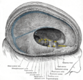 Dura mater and its processes exposed by removing part of the right half of the skull, and the brain.
 -
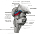
 Superficial dissection of brain-stem. Ventral view.
 -
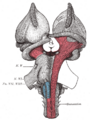
 Hind- and mid-brains; postero-lateral view.
 -
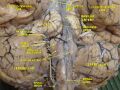 Cerebrum.Inferior view.Deep dissection


External links[edit]
- Animations of extraocular cranial nerve and muscle function and damage (University of Liverpool)
- cranialnerves at The Anatomy Lesson by Wesley Norman (Georgetown University)
(VI
)
| The cranial nerves | ||||||||||
|---|---|---|---|---|---|---|---|---|---|---|
|
Gray's Anatomy[edit]
- Gray's Anatomy Contents
- Gray's Anatomy Subject Index
- About Classic Gray's Anatomy
- Glossary of anatomy terms
Anatomy atlases (external)[edit]
[1] - Anatomy Atlases
| Human systems and organs | ||||||||||||||
|---|---|---|---|---|---|---|---|---|---|---|---|---|---|---|
|

Medical Disclaimer: WikiMD is for informational purposes only and is not a substitute for professional medical advice. Content may be inaccurate or outdated and should not be used for diagnosis or treatment. Always consult your healthcare provider for medical decisions. Verify information with trusted sources such as CDC.gov and NIH.gov. By using this site, you agree that WikiMD is not liable for any outcomes related to its content. See full disclaimer.
Credits:Most images are courtesy of Wikimedia commons, and templates, categories Wikipedia, licensed under CC BY SA or similar.
Translate page: - East Asian
中文,
日本,
한국어,
South Asian
हिन्दी,
தமிழ்,
తెలుగు,
Urdu,
ಕನ್ನಡ,
Southeast Asian
Indonesian,
Vietnamese,
Thai,
မြန်မာဘာသာ,
বাংলা
European
español,
Deutsch,
français,
Greek,
português do Brasil,
polski,
română,
русский,
Nederlands,
norsk,
svenska,
suomi,
Italian
Middle Eastern & African
عربى,
Turkish,
Persian,
Hebrew,
Afrikaans,
isiZulu,
Kiswahili,
Other
Bulgarian,
Hungarian,
Czech,
Swedish,
മലയാളം,
मराठी,
ਪੰਜਾਬੀ,
ગુજરાતી,
Portuguese,
Ukrainian
