Gigantiform cementoma
| Gigantiform cementoma | |
|---|---|
| Synonyms | |
| Pronounce | N/A |
| Specialty | N/A |
| Symptoms | Painless, slow-growing masses in the jaw |
| Complications | Facial deformity, difficulty in chewing |
| Onset | Childhood or adolescence |
| Duration | Chronic |
| Types | N/A |
| Causes | Genetic mutation |
| Risks | Family history |
| Diagnosis | Clinical examination, radiographic imaging, histopathological analysis |
| Differential diagnosis | Fibrous dysplasia, Ossifying fibroma, Cemento-ossifying fibroma |
| Prevention | N/A |
| Treatment | Surgical resection |
| Medication | N/A |
| Prognosis | Good with treatment |
| Frequency | Rare |
| Deaths | N/A |
Gigantiform cementoma is a rare benign fibro-osseous lesion that primarily affects the jaws. It is characterized by the formation of large, painless masses in the jawbones, typically the mandible and less frequently the maxilla. This condition is most commonly diagnosed in children and adolescents, and it has a strong genetic component, often running in families.
Etiology[edit]
Gigantiform cementoma is believed to be caused by a genetic mutation, although the specific gene involved has not been definitively identified. The condition is inherited in an autosomal dominant pattern, meaning that a single copy of the mutated gene from an affected parent can cause the disorder in offspring.
Clinical Presentation[edit]
Patients with gigantiform cementoma typically present with slow-growing, painless masses in the jaw. These masses can lead to significant facial deformity if left untreated. The condition does not usually cause pain or inflammation, but it can result in malocclusion and difficulty in chewing due to the expansion of the jawbones.
Diagnosis[edit]
The diagnosis of gigantiform cementoma is based on a combination of clinical examination, radiographic imaging, and histopathological analysis.
Clinical Examination[edit]
During a clinical examination, the dentist or oral surgeon will assess the size, location, and characteristics of the jaw masses. The absence of pain and the slow growth of the lesions are key clinical features.
Radiographic Imaging[edit]
Radiographic imaging, such as panoramic radiography or computed tomography (CT) scans, is used to evaluate the extent of the lesions. The images typically show well-defined, radiopaque masses within the jawbones, which may be surrounded by a radiolucent rim.
Histopathological Analysis[edit]
A biopsy of the lesion may be performed to confirm the diagnosis. Histopathological analysis reveals a fibro-osseous lesion with cementum-like material and fibrous stroma.
Differential Diagnosis[edit]
The differential diagnosis for gigantiform cementoma includes other fibro-osseous lesions such as:
These conditions can have similar clinical and radiographic features, so careful evaluation is necessary to distinguish them.
Treatment[edit]
The primary treatment for gigantiform cementoma is surgical resection of the affected areas. The goal of surgery is to remove the masses and restore normal jaw function and appearance. In some cases, reconstructive surgery may be necessary to address facial deformities.
Prognosis[edit]
The prognosis for patients with gigantiform cementoma is generally good following surgical treatment. Recurrence is rare, and most patients achieve satisfactory functional and aesthetic outcomes.
Epidemiology[edit]
Gigantiform cementoma is a rare condition, with only a limited number of cases reported in the medical literature. It affects both males and females, and there is no known racial predilection.
See Also[edit]
External Links[edit]
- [Link to relevant medical resources]
Template:Oral and maxillofacial pathology
Gigantiform_cementoma[edit]
-
 Progressive migrating tumours, presented as swollen manifest
 -
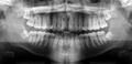 Basic panoramic radiograph
 -
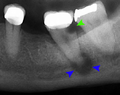 Tooth decay and abscess x-ray


Medical Disclaimer: WikiMD is for informational purposes only and is not a substitute for professional medical advice. Content may be inaccurate or outdated and should not be used for diagnosis or treatment. Always consult your healthcare provider for medical decisions. Verify information with trusted sources such as CDC.gov and NIH.gov. By using this site, you agree that WikiMD is not liable for any outcomes related to its content. See full disclaimer.
Credits:Most images are courtesy of Wikimedia commons, and templates, categories Wikipedia, licensed under CC BY SA or similar.
Translate page: - East Asian
中文,
日本,
한국어,
South Asian
हिन्दी,
தமிழ்,
తెలుగు,
Urdu,
ಕನ್ನಡ,
Southeast Asian
Indonesian,
Vietnamese,
Thai,
မြန်မာဘာသာ,
বাংলা
European
español,
Deutsch,
français,
Greek,
português do Brasil,
polski,
română,
русский,
Nederlands,
norsk,
svenska,
suomi,
Italian
Middle Eastern & African
عربى,
Turkish,
Persian,
Hebrew,
Afrikaans,
isiZulu,
Kiswahili,
Other
Bulgarian,
Hungarian,
Czech,
Swedish,
മലയാളം,
मराठी,
ਪੰਜਾਬੀ,
ગુજરાતી,
Portuguese,
Ukrainian
