The Lumbar Vertebræ
Anatomy > Gray's Anatomy of the Human Body > II. [Osteology]] > The lumbar vertebrae.
Lumbar vertebrae[edit]
General characteristics[edit]

The figure on the left depicts the general characteristics of the first through fourth lumbar vertebrae. The fifth vertebra contains certain peculiarities, which are detailed below.
As with other vertebrae, each lumbar vertebra consists of a vertebral body and a vertebral arch. The vertebral arch, consisting of a pair of pedicles and a pair of laminae, encloses the vertebral foramen (opening) and supports seven processes.
Body[edit]
The vertebral body of each lumbar vertebra is large, wider from side to side than from front to back, and a little thicker in front than in back. It is flattened or slightly concave above and below, concave behind, and deeply constricted in front and at the sides.[1]
Arch[edit]
The pedicles are very strong, directed backward from the upper part of the vertebral body; consequently, the inferior vertebral notches are of considerable depth.[1] The pedicles change in morphology from the upper lumbar to the lower lumbar. They increase in sagittal width from 9 mm to up to 18 mm at L5. They increase in angulation in the axial plane from 10 degrees to 20 degrees by L5. The pedicle is sometimes used as a portal of entrance into the vertebral body for fixation with pedicle screws or for placement of bone cement as with kyphoplasty or vertebroplasty.
The laminae are broad, short, and strong.[1] They form the posterior portion of the vertebral arch. In the upper lumbar region the lamina are taller than wide but in the lower lumbar vertebra the lamina are wider than tall. The lamina connects the spinous process to the pedicles.
The vertebral foramen within the arch is triangular, larger than the thoracic vertebrae, but smaller than in the cervical vertebrae.[1]
Processes[edit]
The spinous process is thick, broad, and somewhat quadrilateral; it projects backward and ends in a rough, uneven border, thickest below where it is occasionally notched.[1]
The superior and inferior articular processes are well-defined, projecting respectively upward and downward from the junctions of pedicles and laminae. The facets on the superior processes are concave, and look backward and medialward; those on the inferior are convex, and are directed forward and lateralward. The former are wider apart than the latter since in the articulated column, the inferior articular processes are embraced by the superior processes of the subjacent vertebra.[1]
The transverse processes are long and slender. They are horizontal in the upper three lumbar vertebrae and incline a little upward in the lower two. In the upper three vertebrae they arise from the junctions of the pedicles and laminae, but in the lower two they are set farther forward and spring from the pedicles and posterior parts of the vertebral bodies. They are situated in front of the articular processes instead of behind them as in the thoracic vertebrae, and are homologous with the ribs.[1]
Three portions or tubercles can be noticed in a transverse process of a lower lumbar vertebrae: the lateral or costiform process, the mammillary process, and the accessory process.[2] The costiform is lateral, the mammillary is superior (cranial), and the accessory is inferior (caudal). The mammillary is connected in the lumbar region with the back part of the superior articular process. [clarification needed]
The accessory process is situated at the back part of the base of the transverse process. The tallest and thickest costiform process is usually that of L5.[2]
First and fifth lumbar vertebrae[edit]

The first lumbar vertebra is level with the anterior end of the ninth rib. This level is also called the important transpyloric plane, since the pylorus of the stomach is at this level. Other important structures are also located at this level, they include; fundus of the gall bladder, celiac trunk, superior mesenteric artery, termination of spinal cord, beginning of filum terminalis, renal vessels, middle suprarenal arteries, and hila of kidneys.
The fifth lumbar vertebra is characterized by its body being much deeper in front than behind, which accords with the prominence of the sacrovertebral articulation; by the smaller size of its spinous process; by the wide interval between the inferior articular processes, and by the thickness of its transverse processes, which spring from the body as well as from the pedicles.[1] The fifth lumbar vertebra is by far the most common site of spondylolysis and spondylolisthesis.[3]
Most individuals have five lumbar vertebrae, while some have four or six. Lumbar disorders that normally affect L5 will affect L4 or L6 in these latter individuals.
Segmental movements[edit]
The range of segmental movements in a single segment is difficult to measure clinically, not only because of variations between individuals, but also because it is age and gender dependent. Furthermore, flexion and extension in the lumbal spine is the product of a combination of rotation and translation in the sagittal plane between each vertebra.[4]
Ranges of segmental movements in the lumbar spine (White and Punjabi, 1990) are (in degrees): [5]
| L1-L2 | L2-L3 | L3-L4 | L4-L5 | L5-S1 | |
|---|---|---|---|---|---|
| Flexion/ Extension |
12° | 14° | 15° | 16° | 17° |
| Lateral flexion |
6° | 6° | 8° | 6° | 3° |
| Axial rotation |
2° | 2° | 2° | 2° | 1° |
Congenital anomalies[edit]
Congenital vertebral anomalies can cause compression of the spinal cord by deforming the vertebral canal or causing instability.
-
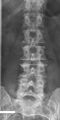 Lumbarization of sacral vertebra 1, seen as 6 vertebrae that do not connect to ribs.
 -
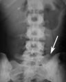 Sacralization of the L5 vertebra is seen at the lower right of the image.
 -
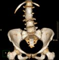 Congenital block vertebra of the lumbar spine. CT volume rendering.


External links[edit]
- Lower Back Pain Condition, Treatment and Exercise(link). SpineUniverse.
- Virtual Spine — Online Learning Resource(link). Toronto Western Hospital Department of Anesthesia and Pain Management.
| Bones of the torso | ||||||||||||||||
|---|---|---|---|---|---|---|---|---|---|---|---|---|---|---|---|---|
|
| Spinal nerves | ||||||||||
|---|---|---|---|---|---|---|---|---|---|---|
|
- ↑ 1.0 1.1 1.2 1.3 1.4 1.5 1.6 1.7 Gray's Anatomy (1918), see infobox
- ↑ 2.0 2.1 Postacchini, Franco (1999) Lumbar Disc Herniation p.19
- ↑ Eizenberg, N. et al. (2008). General Anatomy: Principles and Applications, p. 17.
- ↑ Hansen.Anatomy and Biomechanics of the Back Muscles in the Lumbar Spine With Reference to Biomechanical Modeling(link). Medscape.
- ↑ Hansen.Ranges of Segmental Motion for the Lumbar Spine(link). Medscape.

Medical Disclaimer: WikiMD is for informational purposes only and is not a substitute for professional medical advice. Content may be inaccurate or outdated and should not be used for diagnosis or treatment. Always consult your healthcare provider for medical decisions. Verify information with trusted sources such as CDC.gov and NIH.gov. By using this site, you agree that WikiMD is not liable for any outcomes related to its content. See full disclaimer.
Credits:Most images are courtesy of Wikimedia commons, and templates, categories Wikipedia, licensed under CC BY SA or similar.
Translate page: - East Asian
中文,
日本,
한국어,
South Asian
हिन्दी,
தமிழ்,
తెలుగు,
Urdu,
ಕನ್ನಡ,
Southeast Asian
Indonesian,
Vietnamese,
Thai,
မြန်မာဘာသာ,
বাংলা
European
español,
Deutsch,
français,
Greek,
português do Brasil,
polski,
română,
русский,
Nederlands,
norsk,
svenska,
suomi,
Italian
Middle Eastern & African
عربى,
Turkish,
Persian,
Hebrew,
Afrikaans,
isiZulu,
Kiswahili,
Other
Bulgarian,
Hungarian,
Czech,
Swedish,
മലയാളം,
मराठी,
ਪੰਜਾਬੀ,
ગુજરાતી,
Portuguese,
Ukrainian
