The Oculomotor Nerve
Anatomy > Gray's Anatomy of the Human Body > IX. Neurology > 5c. The Oculomotor Nerve
Henry Gray (1821–1865). Anatomy of the Human Body. 1918.
The Oculomotor Nerve[edit]
(N. Oculomotorius; Third Nerve)
The oculomotor nerve (Figs. 775, 776, 777) supplies somatic motor fibers to all the ocular muscles, except the Obliquus superior and Rectus lateralis; it also supplies through its connections with the ciliary ganglion, sympathetic motor fibers to the Sphincter pupillae and the Ciliaris muscles.

The fibers of the oculomotor nerve arise from a nucleus which lies in the gray substance of the floor of the cerebral aqueduct and extends in front of the aqueduct for a short distance into the floor of the third ventricle. From this nucleus the fibers pass forward through the tegmentum, the red nucleus, and the medial part of the substantia nigra, forming a series of curves with a lateral convexity, and emerge from the oculomotor sulcus on the medial side of the cerebral peduncle.
The nuclei[edit]
The nucleus of the oculomotor nerve does not consist of a continuous column of cells, but is broken up into a number of smaller nuclei, which are arranged in two groups, anterior and posterior.
Those of the posterior group are six in number, five of which are symmetrical on the two sides of the middle line, while the sixth is centrally placed and is common to the nerves of both sides.
The anterior group consists of two nuclei, an antero-medial and an antero-lateral (Fig. 762). The nucleus of the oculomotor nerve, considered from a physiological standpoint, can be subdivided into several smaller groups of cells, each group controlling a particular muscle.
On emerging from the brain, the nerve is invested with a sheath of pia mater, and enclosed in a prolongation from the arachnoid. It passes between the superior cerebellar and posterior cerebral arteries, and then pierces the dura mater in front of and lateral to the posterior clinoid process, passing between the free and attached borders of the tentorium cerebelli.
It runs along the lateral wall of the cavernous sinus, above the other orbital nerves, receiving in its course one or two filaments from the cavernous plexus of the sympathetic, and a communicating branch from the ophthalmic division of the trigeminal.
It then divides into two branches, which enter the orbit through the superior orbital fissure, between the two heads of the Rectus lateralis. Here the nerve is placed below the trochlear nerve and the frontal and lacrimal branches of the ophthalmic nerve, while the nasociliary nerve is placed between its two rami.
The superior ramus the smaller, passes medialward over the optic nerve, and supplies the Rectus superior and Levator palpebrae superioris.
The inferior ramus the larger, divides into three branches. One passes beneath the optic nerve to the Rectus medialis; another, to the Rectus inferior; the third and longest runs forward between the Recti inferior and lateralis to the Obliquus inferior. From the last a short thick branch is given off to the lower part of the ciliary ganglion, and forms its short root All these branches enter the muscles on their ocular surfaces, with the exception of the nerve to the Obliquus inferior, which enters the muscle at its posterior border.
Function[edit]
The oculomotor nerve include axons of type GSE, general somatic efferent, which innervate skeletal muscle of the levator palpebrae superioris, superior rectus, medial rectus, inferior rectus, and inferior oblique muscles.(innervates all the extrinsic muscles except superior oblique and lateral rectus.)
The nerve also includes axons of type GVE, general visceral efferent, which provide preganglionic parasympathetics to the ciliary ganglion. From the ciliary ganglion post ganglionic fibers pass through the short ciliary nerve to the constrictor pupillae of the iris and the cilliary muscles.
Additional images[edit]
-
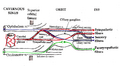
 Map of the oculomotor nerve.
 -
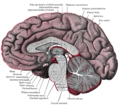 Median sagittal section of brain.
 -
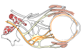
 Pathways in the Ciliary Ganglion.


External links[edit]
- "Oculomotor nerve palsy"
- Oculomotor+Nerve at the US National Library of Medicine Medical Subject Headings (MeSH)
- Animations of extraocular cranial nerve and muscle function and damage (University of Liverpool)
- cranialnerves at The Anatomy Lesson by Wesley Norman (Georgetown University)
(III
)
| The cranial nerves | ||||||||||
|---|---|---|---|---|---|---|---|---|---|---|
|
Gray's Anatomy[edit]
- Gray's Anatomy Contents
- Gray's Anatomy Subject Index
- About Classic Gray's Anatomy
- Glossary of anatomy terms
Anatomy atlases (external)[edit]
[1] - Anatomy Atlases
| Human systems and organs | ||||||||||||||
|---|---|---|---|---|---|---|---|---|---|---|---|---|---|---|
|

Medical Disclaimer: WikiMD is for informational purposes only and is not a substitute for professional medical advice. Content may be inaccurate or outdated and should not be used for diagnosis or treatment. Always consult your healthcare provider for medical decisions. Verify information with trusted sources such as CDC.gov and NIH.gov. By using this site, you agree that WikiMD is not liable for any outcomes related to its content. See full disclaimer.
Credits:Most images are courtesy of Wikimedia commons, and templates, categories Wikipedia, licensed under CC BY SA or similar.
Translate page: - East Asian
中文,
日本,
한국어,
South Asian
हिन्दी,
தமிழ்,
తెలుగు,
Urdu,
ಕನ್ನಡ,
Southeast Asian
Indonesian,
Vietnamese,
Thai,
မြန်မာဘာသာ,
বাংলা
European
español,
Deutsch,
français,
Greek,
português do Brasil,
polski,
română,
русский,
Nederlands,
norsk,
svenska,
suomi,
Italian
Middle Eastern & African
عربى,
Turkish,
Persian,
Hebrew,
Afrikaans,
isiZulu,
Kiswahili,
Other
Bulgarian,
Hungarian,
Czech,
Swedish,
മലയാളം,
मराठी,
ਪੰਜਾਬੀ,
ગુજરાતી,
Portuguese,
Ukrainian

_(14578658070).jpg)