Tongue
The tongue is the fleshy muscle inside the mouth. A tongue lets us taste because the top of the tongue is made mostly of taste buds. It also helps the process of mastication by mixing food with saliva. It is very flexible, so it also helps us eat and talk. The tongue is the strongest muscle in the human body.[1]
Tongue rolling[edit]

Some people can roll their tongue into a tube. The reason why some people are able to and some are not is because of genetic inheritance, meaning that it is based on whether their parents are able to do it. Many schools use tongue rolling as an example of a genetic trait.
People who can roll their tongue can sometimes make a high pitched sound by blowing through their rolled tongue.


External Features[edit]
The tongue exhibits the following external features:
Root[edit]
The root of the tongue is attached to the mandible and hyoid bone by muscles. It is because of these attachments that the tongue is not swallowed during deglutition. The nerve and vessels of the tongue enter through its root.
Tip[edit]
It is the anterior free end of the tongue, which comes into contact with the central incisors.
Body[edit]
The bulk of tongue between the root and tip is called body. It has dorsal and ventral surfaces and right and left lateral margins.
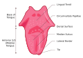
Dorsal surface[edit]
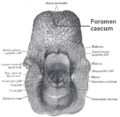
The dorsal surface is convex on all the sides. It is divided by a V-shaped sulcus, the sulcus terminalis into two parts, viz.
- Anterior two-third or oral part.
- Posterior one-third or pharyngeal part.
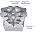
The apex of the sulcus terminalis is marked by a blind foramen, the foramen caecum, which indicates the point of origin of the median thyroid diverticulum (thyroglossal duct) in the embryonic life. The features differ markedly in the oral and pharyngeal parts. The oral part presents the following features:
- A median furrow, representing the bilateral origin of the tongue.
- Large number of papillae.
The pharyngeal part presents the following features:
- A large number of lymphoid follicles, which together constitute the lingual tonsil.
- A large number of mucus and serous glands.
Oral part[edit]
The dorsum of oral part presents a shallow median furrow/groove. The mucus membrane is moist and pink and appears velvety due to the presence of numerous papillae.
- Papillae of the tongue (Lingual papillae): They are projections of lamina propria (corium) of mucus membrane covered with epithelium. The following four chief types of papillae are found:
- Vallate papillae: The vallate papillae (known formerly as circumvallate papillae) are largest (1–2 mm in diameter) and vary in number from 8–12 and are arranged in a V-shaped row in front of sulcus terminalis. Each papilla is like a truncated cone surrounded by a circular sulcus, which is bounded on its periphery by a wall or vallum. The duct of serous glands open into the sulcus (moat) and taste buds are found in the papilla and its vallum.
- Filiform papillae: These are narrowest and most numerous. They are minute conical projections with sharply pointed tips. Filiform papillae are located abundantly on the dorsum of tongue and are largely responsible for its velvety appearance.
- Fungiform papillae: They have a red rounded head (about 1 mm in diameter) and a narrower base, mostly at the apex and margins of the tongue, while some are scattered over the dorsum of the tongue. They are visible as discrete pink pinheads.
- Foliate papillae: They consist of inconstant vertical grooves and ridges near the margin in front of sulcus terminalis. Foliate papillae are more prominent in the tongue of rabbits. They are rudimentary in humans.

Pharyngeal part[edit]
The dorsum of pharyngeal part faces posteriorly and forms the base of tongue. The base of tongue constitutes the anterior wall of the oropharynx and can be inspected only by the use of a mirror or by a downward pressure on the tongue with a tongue spatula. The mucus membrane over the dorsum of pharyngeal part is devoid of papillae. It, however, appears uneven due to the presence of numerous lymphatic follicles in the underlying submucosa. These follicles are collectively termed lingual tonsil.
The mucus membrane in this part is continuous with mucus membrane covering the palatine tonsils and the pharynx. Posteriorly, it is reflected onto the front of the epiglottis as the median glossoepiglottic fold and onto the lateral wall of pharynx as lateral glossoepiglottic folds. The space on each side of the median glossoepiglottic fold is termed epiglottic vallecula.
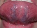
Ventral (inferior) surface of the tongue[edit]
The inferior surface of tongue is situated in the oral cavity only. The mucus membrane lining this surface is thin, smooth, and purplish. It is reflected onto the floor of the mouth. The under aspect of the tongue presents the following features:
- Frenulum linguae, a median-fold of mucus membrane connecting the tongue to the floor of the mouth.
- Deep lingual veins, may be seen through mucous membrane on either side of frenulum linguae (the lingual nerve and lingual artery are medial to the vein but not visible).
- Plica fimbriata, a fringed fimbriated fold of mucous membrane lateral to the lingual vein directed forwards and medially towards the tip of the tongue.
Muscles of the Tongue[edit]
The musculature of tongue consists of extrinsic and intrinsic muscles. The intrinsic muscles are within the tongue and have no attachment outside the tongue whereas extrinsic muscles take origin from structures outside the tongue and enter the tongue to be inserted in it. The intrinsic muscles change the shape of tongue whereas extrinsic muscles move the tongue (such as protrusion, retraction and side-to-side movements) as well as alter its shape.
The tongue is divided into symmetrical right and left halves by a medial fibrous septum, which separates the muscles of two sides. Each half of the tongue contains four intrinsic and four extrinsic muscles. These are as follows:
Intrinsic muscles[edit]
They are confined to the tongue and are not attached to the bone. They occupy the upper part of the tongue and alter its shape. The intrinsic muscles are arranged in several planes. They run in three directions: longitudinal, horizontal, and vertical. The complex interlacing of fibers of these muscles is responsible for the astonishing way in which the tongue can change its shape, becoming wide and flat, narrow and thick, or rolled up laterally to become gutter shaped. The latter shape cannot be achieved by a small number of people and this inability is genetically determined. Intrinsic muscles occupy the upper part of the tongue and are attached to the submucous fibrous layer and to the median fibrous septum.
| Intrinsic
muscle |
Location | Actions |
|---|---|---|
| Superior
longitudinal |
Beneath the
mucous membrane |
|
| Inferior
longitudinal |
Close to inferior
surface between genioglossus and hyoglossus |
|
| Transverse | Extends from
median septum to the margin |
Makes the tongue narrow and elongated |
| Vertical | At the border of
the anterior part of the tongue |
Makes the tongue broad and flattened |
Extrinsic muscles[edit]
They attach the tongue to the mandible (genioglossus), the hyoid (hyoglossus), the styloid process (styloglossus), and the palate (palatoglossus) on each side.

| Muscle | Origin | Insertion | Actions |
|---|---|---|---|
| Genioglossus (a fanshaped
muscle) |
Superior genial tubercle |
|
Protrudes the tongue when acting
together with its counterpart of opposite side |
| Hyoglossus (a flat
quadrilateral muscle) |
Greater cornu and adjacent
part of the body of hyoid |
Side of tongue (posterior half) between
styloglossus laterally and inferior longitudinal muscle medially |
|
| Styloglossus (an
elongated slip) |
Tip of styloid process and
adjacent part of the stylohyoid ligament |
Side of tongue (whole length),
interdigitating posteriorly with the fibres of hyoglossus |
Draws the side of the tongue upwards
and backwards |
| Palatoglossus (a slender
slip) |
Oral surface of palatine
aponeurosis of palate |
Side of tongue (at the junction of its
oral and pharyngeal parts) |
|
Blood Supply[edit]
Arterial Supply[edit]
The tongue is supplied by the following arteries:
- Branches of lingual artery (chief artery of tongue); the deep lingual arteries to the anterior part and dorsal lingual arteries to the posterior part.
- Tonsillar branch of the facial artery.
- Ascending pharyngeal artery.

Venous Drainage[edit]
It is by the following veins:
- Deep lingual vein is the principal vein of the tongue and is visible on the inferior surface of the tongue near the median plane through thin mucous membrane in life.
- Venae comitantes accompanying the lingual artery. They are joined by dorsal lingual veins.
- Venae comitantes accompanying the hypoglossal nerve. These veins unite at the posterior border of the hyoglossus to form the lingual vein, which drains into either common facial vein or internal jugular vein.
Lymphatic Drainage[edit]
The lymphatics emerging from the tongue are grouped into the following four sets:
- Apical vessels: They drain the tip and inferior surface of the tongue into submental lymph nodes after piercing the mylohyoid muscle. Their efferents go to the submandibular nodes mainly, some cross the hyoid bone to reach the jugulo-omohyoid nodes.
- Marginal vessels: They drain the marginal portions of the anterior two-third of the tongue—unilaterally into submandibular lymph nodes and then to the lower deep cervical lymph nodes, including jugulo-omohyoid.
- Central vessels: They drain the central portion of the anterior two-third of the tongue (i.e., area within 0.5 inch on either side of midline). They pass vertically downwards in the midline of the tongue between the genioglossus muscles and then drain bilaterally into the deep cervical lymph nodes.
- Basal vessels: They drain the root of the tongue and posterior one-third of the tongue bilaterally into upper deep cervical lymph nodes, including jugulodigastric.
Nerve Supply[edit]
The nerves supplying the tongue are as follows:
- Motor supply: All the muscles of tongue (intrinsic and extrinsic) are supplied by the hypoglossal nerve except palatoglossus which is supplied by cranial root of accessory via pharyngeal plexus.
- Sensory supply:
Anterior two-third of the tongue is supplied by:
- lingual nerve carrying general sensations, and
- chorda tympani nerve carrying special sensations of taste.
Posterior one-third of the tongue is supplied by:
- glossopharyngeal nerve, carrying both general and special sensations of taste, and
- posteriormost part (base of the tongue), supplied by the internal laryngeal branch of the superior laryngeal carrying special sensations of taste.
Nerves carrying taste sensations from the tongue are as follows:
- Chorda tympani nerve (a branch of the facial nerve) from anterior two-third of the tongue.
- Glossopharyngeal from posterior one-third of the tongue.
- Internal laryngeal nerve from superior laryngeal branch of the vagus nerve, from posteriormost part of the tongue.
Gallery[edit]
-
 Tongue
 -
 Facies inferior linguae
 -
 Foramen caecum
 -
 Tongue
 -
 Gray's Anatomy illustration
 -
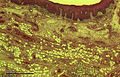 Human tongue section
 -
 Gray's Anatomy illustration
 -
 Tongue and taste buds


_Section.jpg)


| Anatomy of the gastrointestinal tract, excluding the mouth | ||||||||||||
|---|---|---|---|---|---|---|---|---|---|---|---|---|
|

- ↑ interesting tongue fact at Strange Facts.com

Medical Disclaimer: WikiMD is for informational purposes only and is not a substitute for professional medical advice. Content may be inaccurate or outdated and should not be used for diagnosis or treatment. Always consult your healthcare provider for medical decisions. Verify information with trusted sources such as CDC.gov and NIH.gov. By using this site, you agree that WikiMD is not liable for any outcomes related to its content. See full disclaimer.
Credits:Most images are courtesy of Wikimedia commons, and templates, categories Wikipedia, licensed under CC BY SA or similar.
Translate page: - East Asian
中文,
日本,
한국어,
South Asian
हिन्दी,
தமிழ்,
తెలుగు,
Urdu,
ಕನ್ನಡ,
Southeast Asian
Indonesian,
Vietnamese,
Thai,
မြန်မာဘာသာ,
বাংলা
European
español,
Deutsch,
français,
Greek,
português do Brasil,
polski,
română,
русский,
Nederlands,
norsk,
svenska,
suomi,
Italian
Middle Eastern & African
عربى,
Turkish,
Persian,
Hebrew,
Afrikaans,
isiZulu,
Kiswahili,
Other
Bulgarian,
Hungarian,
Czech,
Swedish,
മലയാളം,
मराठी,
ਪੰਜਾਬੀ,
ગુજરાતી,
Portuguese,
Ukrainian
