Aponeurosis of the abdominal external oblique muscle
-
 Aponeurosis of the abdominal external oblique muscle
 -
 Aponeurosis of the abdominal external oblique muscle


Aponeurosis of the Abdominal External Oblique Muscle[edit]
The aponeurosis of the abdominal external oblique muscle is a broad, flat tendon that plays a crucial role in the structure and function of the abdominal wall. It is part of the muscular system and contributes to the anterior abdominal wall's strength and flexibility.
Structure[edit]
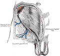
The aponeurosis of the external oblique muscle is a fibrous sheet that extends from the lower ribs to the linea alba, the pubic tubercle, and the iliac crest. It is formed by the tendinous fibers of the external oblique muscle, which is the largest and most superficial of the three flat muscles of the lateral anterior abdomen.
The aponeurosis is composed of dense connective tissue and is characterized by its shiny, white appearance. It is continuous with the muscle fibers of the external oblique, which originate from the lower eight ribs and interdigitate with the serratus anterior and latissimus dorsi muscles.
Function[edit]
The primary function of the aponeurosis of the external oblique is to provide a strong, flexible support for the abdominal wall. It helps to:
- Maintain the integrity of the abdominal cavity, protecting the internal organs.
- Assist in movements such as flexion, lateral flexion, and rotation of the trunk.
- Contribute to the Valsalva maneuver, which is important for activities such as defecation, urination, and childbirth.
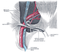
The aponeurosis also plays a role in the formation of the inguinal ligament, which is formed by the lower border of the aponeurosis as it folds back on itself.
Clinical Significance[edit]
The aponeurosis of the external oblique is clinically significant in several ways:
- It is involved in the formation of the inguinal canal, a potential site for inguinal hernias.
- Surgical incisions in the abdominal wall often involve cutting through or around the aponeurosis, requiring careful consideration to avoid weakening the abdominal wall.
- Injuries or tears to the aponeurosis can result from trauma or overuse, leading to pain and dysfunction.
Related Structures[edit]
The aponeurosis of the external oblique is related to several other structures in the abdominal region:
- The internal oblique muscle and transversus abdominis muscle, which lie deep to the external oblique and have their own aponeuroses.
- The rectus abdominis muscle, which is enclosed in the rectus sheath formed by the aponeuroses of the abdominal muscles.
- The inguinal ligament, which is formed by the lower edge of the aponeurosis.
Related Pages[edit]
| Anatomy and morphology | ||||||||||
|---|---|---|---|---|---|---|---|---|---|---|
|

Medical Disclaimer: WikiMD is for informational purposes only and is not a substitute for professional medical advice. Content may be inaccurate or outdated and should not be used for diagnosis or treatment. Always consult your healthcare provider for medical decisions. Verify information with trusted sources such as CDC.gov and NIH.gov. By using this site, you agree that WikiMD is not liable for any outcomes related to its content. See full disclaimer.
Credits:Most images are courtesy of Wikimedia commons, and templates, categories Wikipedia, licensed under CC BY SA or similar.
Translate page: - East Asian
中文,
日本,
한국어,
South Asian
हिन्दी,
தமிழ்,
తెలుగు,
Urdu,
ಕನ್ನಡ,
Southeast Asian
Indonesian,
Vietnamese,
Thai,
မြန်မာဘာသာ,
বাংলা
European
español,
Deutsch,
français,
Greek,
português do Brasil,
polski,
română,
русский,
Nederlands,
norsk,
svenska,
suomi,
Italian
Middle Eastern & African
عربى,
Turkish,
Persian,
Hebrew,
Afrikaans,
isiZulu,
Kiswahili,
Other
Bulgarian,
Hungarian,
Czech,
Swedish,
മലയാളം,
मराठी,
ਪੰਜਾਬੀ,
ગુજરાતી,
Portuguese,
Ukrainian
