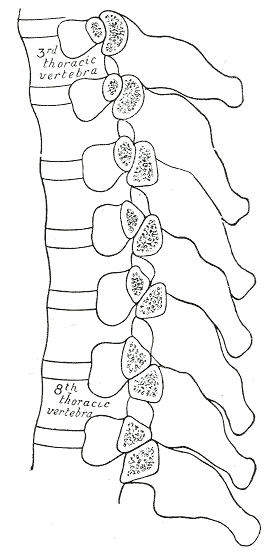
Costovertebral Articulations
Editor-In-Chief: Prab R Tumpati, MD
Obesity, Sleep & Internal medicine
Founder, WikiMD Wellnesspedia &
W8MD medical weight loss NYC and sleep center NYC
Anatomy > Gray's Anatomy of the Human Body > Syndesmology > Costovertebral Articulations
Henry Gray (1821–1865). Anatomy of the Human Body. 1918.
Costovertebral Articulations[edit | edit source]
(Articulationes Costovertebrales)
The costovertebral articulations are the joints between the ribs and the vertebral column. These are divided into two main types:
- The costocentral articulations: between the head of the rib and the vertebral bodies.
- The costotransverse articulations: between the tubercle of the rib and the transverse processes of the vertebrae.
Articulations of the Heads of the Ribs (articulationes capitulorum costarum)[edit | edit source]
These are gliding (arthrodial) joints formed between the head of the rib and:
- The demi-facets on the contiguous vertebral bodies.
- The intervening intervertebral disc.
Note: The 1st, 10th, 11th, and 12th ribs typically articulate with a single vertebra, while ribs 2–9 articulate with two adjacent vertebrae.
Ligaments[edit | edit source]
- Articular capsule – Encloses the joint, connecting the rib head to the vertebral body and disc.
- Radiate ligament (ligamentum capituli costae radiatum) – A triradiate ligament attaching the rib head to the vertebral bodies and the disc.
- Interarticular ligament – Connects the ridge between the rib head's articular facets to the intervertebral disc, dividing the joint into two cavities.
The radiate ligament is especially important anteriorly, while the interarticular ligament is absent in the joints of the 1st, 10th, 11th, and 12th ribs.
Synovial Membranes[edit | edit source]
- Ribs with two articular facets (ribs 2–9) have two synovial cavities.
- Ribs with a single facet (1st, 10th–12th) have one synovial cavity.
Costotransverse Articulations (articulationes costotransversariae)[edit | edit source]
These joints connect the articular portion of the rib's tubercle to the transverse process of the corresponding vertebra. Absent in the 11th and 12th ribs.
Ligaments of the Costotransverse Joint[edit | edit source]
- Articular capsule – Surrounds the synovial joint between the rib tubercle and transverse process.
- Anterior costotransverse ligament – Extends from the neck of the rib to the transverse process above.
- Posterior costotransverse ligament – Connects the neck of the rib to the base of the transverse process and the inferior articular process of the vertebra above.
- Ligament of the neck of the rib – A strong band between the posterior rib neck and the anterior transverse process.
- Ligament of the tubercle of the rib – Connects the rough non-articular part of the tubercle to the transverse process apex.
Associated Ligaments and Structures[edit | edit source]
- The first rib lacks an anterior costotransverse ligament.
- The twelfth rib connects to the transverse process of the first lumbar vertebra via the lumbocostal ligament (a continuation of the thoracolumbar fascia).
Movements of the Costovertebral Joints[edit | edit source]
The nature of movement at these joints is determined by:
- The shape of the joint surfaces.
- The arrangement of the ligaments.
Upper Ribs (1–6)[edit | edit source]
- Rotation around the long axis of the neck of the rib.
- Elevation → anterior rotation; Depression → posterior rotation.
- Tubercle moves within a concave facet on the anterior transverse process.
Lower Ribs (7–10)[edit | edit source]
- The articular surfaces are flat and oriented inferiorly, medially, and posteriorly.
- Movements involve gliding: upward/backward/medial or downward/forward/lateral.
- Limited axial rotation accompanies these gliding motions.
Floating Ribs (11–12)[edit | edit source]
- Lack costotransverse joints.
- Increased freedom of motion, particularly in respiration.
The costocentral and costotransverse articulations function in concert, forming a composite joint. The total effect is the rotation and slight elevation or depression of the rib around a central axis.
Clinical Relevance[edit | edit source]
- Thoracic trauma can disrupt these joints, leading to pain and impaired breathing mechanics.
- Costovertebral joint dysfunction may present as localized back pain.
- These joints play a crucial role in the biomechanics of respiration.
See Also[edit | edit source]
- Thoracic vertebrae
- Ribs
- Costal cartilage
- Sternocostal joints
- Articulations of the vertebral column
- Movements of the thorax
- Respiratory system
| Joints and ligaments of torso | ||||||||||||||
|---|---|---|---|---|---|---|---|---|---|---|---|---|---|---|
|
Gray's Anatomy[edit source]
- Gray's Anatomy Contents
- Gray's Anatomy Subject Index
- About Classic Gray's Anatomy
- Glossary of anatomy terms
Anatomy atlases (external)[edit source]
[1] - Anatomy Atlases
| Human systems and organs | ||||||||||||||
|---|---|---|---|---|---|---|---|---|---|---|---|---|---|---|
|
Adapted from the Classic Grays Anatomy of the Human Body 1918 edition (public domain)
Search WikiMD
Ad.Tired of being Overweight? Try W8MD's physician weight loss program.
Semaglutide (Ozempic / Wegovy and Tirzepatide (Mounjaro / Zepbound) available.
Advertise on WikiMD
|
WikiMD's Wellness Encyclopedia |
| Let Food Be Thy Medicine Medicine Thy Food - Hippocrates |
Translate this page: - East Asian
中文,
日本,
한국어,
South Asian
हिन्दी,
தமிழ்,
తెలుగు,
Urdu,
ಕನ್ನಡ,
Southeast Asian
Indonesian,
Vietnamese,
Thai,
မြန်မာဘာသာ,
বাংলা
European
español,
Deutsch,
français,
Greek,
português do Brasil,
polski,
română,
русский,
Nederlands,
norsk,
svenska,
suomi,
Italian
Middle Eastern & African
عربى,
Turkish,
Persian,
Hebrew,
Afrikaans,
isiZulu,
Kiswahili,
Other
Bulgarian,
Hungarian,
Czech,
Swedish,
മലയാളം,
मराठी,
ਪੰਜਾਬੀ,
ગુજરાતી,
Portuguese,
Ukrainian
Medical Disclaimer: WikiMD is not a substitute for professional medical advice. The information on WikiMD is provided as an information resource only, may be incorrect, outdated or misleading, and is not to be used or relied on for any diagnostic or treatment purposes. Please consult your health care provider before making any healthcare decisions or for guidance about a specific medical condition. WikiMD expressly disclaims responsibility, and shall have no liability, for any damages, loss, injury, or liability whatsoever suffered as a result of your reliance on the information contained in this site. By visiting this site you agree to the foregoing terms and conditions, which may from time to time be changed or supplemented by WikiMD. If you do not agree to the foregoing terms and conditions, you should not enter or use this site. See full disclaimer.
Credits:Most images are courtesy of Wikimedia commons, and templates, categories Wikipedia, licensed under CC BY SA or similar.
Contributors: Anish, Deepika vegiraju, Prab R. Tumpati, MD


