The Lateral Vertebral Muscles
Anatomy > Gray's Anatomy of the Human Body > IV. Myology > 5e. The Lateral Vertebral Muscles
Henry Gray (1821–1865). Anatomy of the Human Body. 1918.
The Lateral Vertebral Muscles[edit]
The lateral vertebral muscles (Fig. 387). are:
Scalenus anterior[edit]
The Scalenus anterior (Scalenus anticus) lies deeply at the side of the neck, behind the Sternocleidomastoideus. It arises from the anterior tubercles of the transverse processes of the third, fourth, fifth, and sixth cervical vertebrae, and descending, almost vertically, is inserted by a narrow, flat tendon into the scalene tubercle on the inner border of the first rib, and into the ridge on the upper surface of the rib in front of the subclavian groove.
Scalenus medius[edit]
The Scalenus medius the largest and longest of the three Scaleni, arises from the posterior tubercles of the transverse processes of the lower six cervical vertebrae, and descending along the side of the vertebral column, is inserted by a broad attachment into the upper surface of the first rib, between the tubercle and the subclavian groove.
Scalenus posterior[edit]
The Scalenus posterior (Scalenus posticus), the smallest and most deeply seated of the three Scaleni, arises by two or three separate tendons, from the posterior tubercles of the transverse processes of the lower two or three cervical vertebrae, and is inserted by a thin tendon into the outer surface of the second rib, behind the attachment of the Serratus anterior. It is occasionally blended with the Scalenus medius.
Variations[edit]
The Scaleni muscles vary considerably in their attachments and in the arrangement of their fibers. A slip from the Scalenus anticus may pass behind the subclavian artery. The Scalenus posticus may be absent or extend to the third rib. The Scalenus pleuralis muscle extends from the transverse process of the seventh cervical vertebra to the fascia supporting the dome of the pleura and inner border of first rib.
Nerves[edit]
The Scaleni are supplied by branches from the second to the seventh cervical nerves.
Actions[edit]
When the Scaleni act from above, they elevate the first and second ribs, and are, therefore, inspiratory muscles. Acting from below, they bend the vertebral column to one or other side; if the muscles of both sides act, the vertebral column is slightly flexed.
Function[edit]
The anterior and middle scalene muscles lifts the first rib and bends the neck to the same side as the acting muscle;[1] the posterior scalene lifts the second rib and tilts the neck to the same side.
Because they elevate the upper ribs they also act as accessory muscles of respiration, along with the sternocleidomastoids.
Additional images[edit]
-
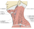 Musculi colli base
 -
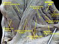 Scalene muscles. Muscles of the neck. Lateral view.
 -
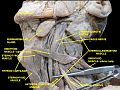 Scalene muscles. Muscles of the neck. Lateral view.
 -
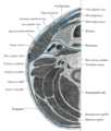 Section of the neck at about the level of the sixth cervical vertebra. Showing the arrangement of the fascia coli.


References[edit]
- ↑ "Actions of the scalene muscles for rotation of the cervical spine in macaque and human".J Orthop Sports Phys Ther.Oct 2002;32(10)
- 488–96.doi:10.2519/jospt.2002.32.10.488.PMID:12403200.
| Muscles of the neck | ||||||||||
|---|---|---|---|---|---|---|---|---|---|---|
|
Gray's Anatomy[edit]
- Gray's Anatomy Contents
- Gray's Anatomy Subject Index
- About Classic Gray's Anatomy
- Glossary of anatomy terms
Anatomy atlases (external)[edit]
[1] - Anatomy Atlases
| Human systems and organs | ||||||||||||||
|---|---|---|---|---|---|---|---|---|---|---|---|---|---|---|
|

Medical Disclaimer: WikiMD is for informational purposes only and is not a substitute for professional medical advice. Content may be inaccurate or outdated and should not be used for diagnosis or treatment. Always consult your healthcare provider for medical decisions. Verify information with trusted sources such as CDC.gov and NIH.gov. By using this site, you agree that WikiMD is not liable for any outcomes related to its content. See full disclaimer.
Credits:Most images are courtesy of Wikimedia commons, and templates, categories Wikipedia, licensed under CC BY SA or similar.
Translate page: - East Asian
中文,
日本,
한국어,
South Asian
हिन्दी,
தமிழ்,
తెలుగు,
Urdu,
ಕನ್ನಡ,
Southeast Asian
Indonesian,
Vietnamese,
Thai,
မြန်မာဘာသာ,
বাংলা
European
español,
Deutsch,
français,
Greek,
português do Brasil,
polski,
română,
русский,
Nederlands,
norsk,
svenska,
suomi,
Italian
Middle Eastern & African
عربى,
Turkish,
Persian,
Hebrew,
Afrikaans,
isiZulu,
Kiswahili,
Other
Bulgarian,
Hungarian,
Czech,
Swedish,
മലയാളം,
मराठी,
ਪੰਜਾਬੀ,
ગુજરાતી,
Portuguese,
Ukrainian
