The Subclavian Artery
Anatomy > Gray's Anatomy of the Human Body > VI. The Arteries > 4. The Arteries of the Upper Extremity. a. The Subclavian Artery
Henry Gray (1821–1865). Anatomy of the Human Body. 1918.
4. The Arteries of the Upper Extremity. a. The Subclavian Artery The artery which supplies the upper extremity continues as a single trunk from its commencement down to the elbow; but different portions of it have received different names, according to the regions through which they pass. That part of the vessel which extends from its origin to the outer border of the first rib is termed the subclavian beyond this point to the lower border of the axilla it is named the axillary and from the lower margin of the axillary space to the bend of the elbow it is termed brachial here the trunk ends by dividing into two branches the radial and ulnar
The Subclavian Artery (A. Subclavia) (Fig. 520) —On the right side the subclavian artery arises from the innominate artery behind the right sternoclavicular articulation; on the left side it springs from the arch of the aorta. The two vessels, therefore, in the first part of their course, differ in length, direction, and relation with neighboring structures. In order to facilitate the description, each subclavian artery is divided into three parts. The first portion extends from the origin of the vessel to the medial border of the Scalenus anterior; the second lies behind this muscle; and the third extends from the lateral margin of the muscle to the outer border of the first rib, where it becomes the axillary artery. The first portions of the two vessels require separate descriptions; the second and third parts of the two arteries are practically alike.
First Part of the Right Subclavian Artery (Figs. 505, 520)—The first part of the right subclavian artery arises from the innominate artery, behind the upper part of the right sternoclavicular articulation, and passes upward and lateralward to the medial margin of the Scalenus anterior. It ascends a little above the clavicle, the extent to which it does so varying in different cases.

Relations—It is covered, in front by the integument, superficial fascia, Platysma, deep fascia, the clavicular origin of the Sternocleidomastoideus, the Sternohyoideus, and Sternothyreoideus, and another layer of the deep fascia. It is crossed by the internal jugular and vertebral veins, by the vagus nerve and the cardiac branches of the vagus and sympathetic, and by the subclavian loop of the sympathetic trunk which forms a ring around the vessel. The anterior jugular vein is directed lateralward in front of the artery, but is separated from it by the Sternohyoideus and Sternothyreoideus. Below and behind the artery is the pleura, which separates it from the apex of the lung; behind is the sympathetic trunk, the Longus collie and the first thoracic vertebra. The right recurrent nerve winds around the lower and back part of the vessel.
First Part of the Left Subclavian Artery (Fig. 505)—The first part of the left subclavian artery arises from the arch of the aorta, behind the left common carotid, and at the level of the fourth thoracic vertebra; it ascends in the superior mediastinal cavity to the root of the neck and then arches lateralward to the medial border of the Scalenus anterior.
Relations—It is in relation, in front with the vagus, cardiac, and phrenic nerves, which lie parallel with it, the left common carotid artery, left internal jugular and vertebral veins, and the commencement of the left innominate vein, and is covered by the Sternothyreoideus, Sternohyoideus, and Sternocleidomastoideus; behind it is in relation with the esophagus, thoracic duct, left recurrent nerve, inferior cervical ganglion of the sympathetic trunk, and Longus colli; higher up, however, the esophagus and thoracic duct lie to its right side; the latter ultimately arching over the vessel to join the angle of union between the subclavian and internal jugular veins. Medial to it are the esophagus, trachea, thoracic duct, and left recurrent nerve; lateral to it, the left pleura and lung.
Second and Third Parts of the Subclavian Artery (Fig. 520)—The second portion of the subclavian artery lies behind the Scalenus anterior; it is very short, and forms the highest part of the arch described by the vessel.
Relations—It is covered, in front by the skin, superficial fascia, Platysma, deep cervical fascia, Sternocleidomastoideus, and Scalenus anterior. On the right side of the neck the phrenic nerve is separated from the second part of the artery by the Scalenus anterior, while on the left side it crosses the first part of the artery close to the medial edge of the muscle. Behind the vessel are the pleura and the Scalenus medius; above the brachial plexus of nerves; below the pleura. The subclavian vein lies below and in front of the artery, separated from it by the Scalenus anterior.
The third portion of the subclavian artery runs downward and lateralward from the lateral margin of the Scalenus anterior to the outer border of the first rib, where it becomes the axillary artery. This is the most superficial portion of the vessel, and is contained in the subclavian triangle (see page 565).
Relations—It is covered, in front by the skin, the superficial fascia, the Platysma, the supraclavicular nerves, and the deep cervical fascia. The external jugular vein crosses its medial part and receives the transverse scapular, transverse cervical, and anterior jugular veins, which frequently form a plexus in front of the artery. Behind the veins, the nerve to the Subclavius descends in front of the artery. The terminal part of the artery lies behind the clavicle and the Subclavius and is crossed by the transverse scapular vessels. The subclavian vein is in front of and at a slightly lower level than the artery. Behind it lies on the lowest trunk of the brachial plexus, which intervenes between it and the Scalenus medius. Above and to its lateral side are the upper trunks of the brachial plexus and the Omohyoideus. Below it rests on the upper surface of the first rib.
Peculiarities—The subclavian arteries vary in their origin, their course, and the height to which they rise in the neck.
- The origin of the right subclavian from the innominate takes place, in some cases, above the sternoclavicular articulation, and occasionally, but less frequently, below that joint. The artery may arise as a separate trunk from the arch of the aorta, and in such cases it may be either the first, second, third, or even the last branch derived from that vessel; in the majority, however, it is the first or last, rarely the second or third. When it is the first branch, it occupies the ordinary position of the innominate artery; when the second or third, it gains its usual position by passing behind the right carotid; and when the last branch, it arises from the left extremity of the arch, and passes obliquely toward the right side, usually behind the trachea, esophagus, and right carotid, sometimes between the esophagus and trachea, to the upper border of the first rib, whence it follows its ordinary course. In very rare instances, this vessel arises from the thoracic aorta, as low down as the fourth thoracic vertebra. Occasionally, it perforates the Scalenus anterior; more rarely it passes in front of that muscle. Sometimes the subclavian vein passes with the artery behind the Scalenus anterior. The artery may ascend as high as 4 cm. above the clavicle, or any intermediate point between this and the upper border of the bone, the right subclavian usually ascending higher than the left.
- The left subclavian is occasionally joined at its origin with the left carotid.
- The left subclavian artery is more deeply placed than the right in the first part of its course, and, as a rule, does not reach quite as high a level in the neck. The posterior border of the Sternocleidomastoideus corresponds pretty closely to the lateral border of the Scalenus anterior, so that the third portion of the artery, the part most accessible for operation, lies immediately lateral to the posterior border of the Sternocleidomastoideus.
- Collateral Circulation—After ligature of the third part of the subclavian artery, the collateral circulation is established mainly by three sets of vessels, thus described in a dissection:
- 1. A posterior set, consisting of the transverse scapular and the descending ramus of the transverse cervical branches of the subclavian, anastomosing with the subscapular from the axillary.
- 2. A medial set, produced by the connection of the internal mammary on the one hand, with the highest intercostal and lateral thoracic arteries, and the branches from the subscapular on the other.
- 3. A middle or axillary set, consisting of a number of small vessels derived from branches of the subclavian, above, and, passing through the axilla, terminating either in the main trunk, or some of the branches of the axillary below. This last set presented most conspicuously the peculiar character of newly formed or, rather, dilated arteries, being excessively tortuous, and forming a complete plexus.
- The chief agent in the restoration of the axillary artery below the tumor was the subscapular artery, which communicated most freely with the internal mammary, transverse scapular and descending ramus of the transverse cervical branches of the subclavian, from all of which it received so great an influx of blood as to dilate it to three times its natural size. 101 20
- When a ligature is applied to the first part of the subclavian artery, the collateral circulation is carried on by: (1) the anastomosis between the superior and inferior thyroids; (2) the anastomosis of the two vertebrals; (3) the anastomosis of the internal mammary with the inferior epigastric and the aortic intercostals; (4) the costocervical anastomosing with the aortic intercostals; (5) the profunda cervicis anastomosing with the descending branch of the occipital; (6) the scapular branches of the thyrocervical trunk anastomosing with the branches of the axillary, and (7) the thoracic branches of the axillary anastomosing with the aortic intercostals.
- Branches—The branches of the subclavian artery are:
- Vertebral.
- Internal mammary.
- Thyrocervical.
- Costocervical.
- On the left side all four branches generally arise from the first portion of the vessel; but on the right side (Fig. 520) the costocervical trunk usually springs from the second portion of the vessel. On both sides of the neck, the first three branches arise close together at the medial border of the Scalenus anterior; in the majority of cases, a free interval of from 1.25 to 2.5 cm. exists between the commencement of the artery and the origin of the nearest branch.
- 1. The vertebral artery (a. vertebralis) (Fig. 514), is the first branch of the subclavian, and arises from the upper and back part of the first portion of the vessel. It is surrounded by a plexus of nerve fibers derived from the inferior cervical ganglion of the sympathetic trunk, and ascends through the foramina in the transverse processes of the upper six cervical vertebræ 102 it then winds behind the superior articular process of the atlas and, entering the skull through the foramen magnum, unites, at the lower border of the pons, with the vessel of the opposite side to form the basilar artery.
- Relations—The vertebral artery may be divided into four parts: The first part runs upward and backward between the Longus colli and the Scalenus anterior. In front of it are the internal jugular and vertebral veins, and it is crossed by the inferior thyroid artery; the left vertebral is crossed by the thoracic duct also. Behind it are the transverse process of the seventh cervical vertebra, the sympathetic trunk and its inferior cervical ganglion. The second part runs upward through the foramina in the transverse processes of the upper six cervical vertebræ, and is surrounded by branches from the inferior cervical sympathetic ganglion and by a plexus of veins which unite to form the vertebral vein at the lower part of the neck. It is situated in front of the trunks of the cervical nerves, and pursues an almost vertical course as far as the transverse process of the atlas, above which it runs upward and lateralward to the foramen in the transverse process of the atlas. The third part issues from the latter foramen on the medial side of the Rectus capitis lateralis, and curves backward behind the superior articular process of the atlas, the anterior ramus of the first cervical nerve being on its medial side; it then lies in the groove on the upper surface of the posterior arch of the atlas, and enters the vertebral canal by passing beneath the posterior atlantoöccipital membrane. This part of the artery is covered by the Semispinalis capitis and is contained in the suboccipital triangle—a triangular space bounded by the Rectus capitis posterior major, the Obliquus superior, and the Obliquus inferior. The first cervical or suboccipital nerve lies between the artery and the posterior arch of the atlas. The fourth part pierces the dura mater and inclines medialward to the front of the medulla oblongata; it is placed between the hypoglossal nerve and the anterior root of the first cervical nerve and beneath the first digitation of the ligamentum denticulatum. At the lower border of the pons it unites with the vessel of the opposite side to form the basilar artery.
- Branches—The branches of the vertebral artery may be divided into two sets: those given off in the neck, and those within the cranium.
- Cervical Branches Cranial Branches
- Spinal. Meningeal.
- Muscular. Posterior Spinal.
- Anterior Spinal.
- Posterior Inferior Cerebellar.
- Medullary.
- Spinal Branches (rami spinales) enter the vertebral canal through the intervertebral foramina, and each divides into two branches. Of these, one passes along the roots of the nerves to supply the medulla spinalis and its membranes, anastomosing with the other arteries of the medulla spinalis; the other divides into an ascending and a descending branch, which unite with similar branches from the arteries above and below, so that two lateral anastomotic chains are formed on the posterior surfaces of the bodies of the vertebræ, near the attachment of the pedicles. From these anastomotic chains branches are supplied to the periosteum and the bodies of the vertebræ, and others form communications with similar branches from the opposite side; from these communications small twigs arise which join similar branches above and below, to form a central anastomotic chain on the posterior surface of the bodies of the vertebræ.
- Muscular Branches are given off to the deep muscles of the neck, where the vertebral artery curves around the articular process of the atlas. They anastomose with the occipital, and with the ascending and deep cervical arteries.
- The Meningeal Branch (ramus meningeus; posterior meningeal branch) springs from the vertebral opposite the foramen magnum, ramifies between the bone and dura mater in the cerebellar fossa, and supplies the falx cerebelli. It is frequently represented by one or two small branches.
- The Posterior Spinal Artery (a. spinalis posterior; dorsal spinal artery) arises from the vertebral, at the side of the medulla oblongata; passing backward, it descends on this structure, lying in front of the posterior roots of the spinal nerves, and is reinforced by a succession of small branches, which enter the vertebral canal through the intervertebral foramina; by means of these it is continued to the lower part of the medulla spinalis, and to the cauda equina. Branches from the posterior spinal arteries form a free anastomosis around the posterior roots of the spinal nerves, and communicate, by means of very tortuous transverse branches, with the vessels of the opposite side. Close to its origin each gives off an ascending branch, which ends at the side of the fourth ventricle.
- The Anterior Spinal Artery (a. spinalis anterior; ventral spinal artery) is a small branch, which arises near the termination of the vertebral, and, descending in front of the medulla oblongata, unites with its fellow of the opposite side at the level of the foramen magnum. One of these vessels is usually larger than the other, but occasionally they are about equal in size. The single trunk, thus formed, descends on the front of the medulla spinalis, and is reinforced by a succession of small branches which enter the vertebral canal through the intervertebral foramina; these branches are derived from the vertebral and the ascending cervical of the inferior thyroid in the neck; from the intercostals in the thorax; and from the lumbar, iliolumbar, and lateral sacral arteries in the abdomen and pelvis. They unite, by means of ascending and descending branches, to form a single anterior median artery, which extend as far as the lower part of the medulla spinalis, and is continued as a slender twig on the filum terminale. This vessel is placed in the pia mater along the anterior median fissure; it supplies that membrane, and the substance of the medulla spinalis, and sends off branches at its lower part to be distributed to the cauda equina.
- The Posterior Inferior Cerebellar Artery (a. cerebelli inferior posterior) (Fig. 516), the largest branch of the vertebral, winds backward around the upper part of the medulla oblongata, passing between the origins of the vagus and accessory nerves, over the inferior peduncle to the under surface of the cerebellum, where it divides into two branches. The medial branch is continued backward to the notch between the two hemispheres of the cerebellum; while the lateral supplies the under surface of the cerebellum, as far as its lateral border, where it anastomoses with the anterior inferior cerebellar and the superior cerebellar branches of the basilar artery. Branches from this artery supply the choroid plexus of the fourth ventricle.
- The Medullary Arteries (bulbar arteries) are several minute vessels which spring from the vertebral and its branches and are distributed to the medulla oblongata.
- The Basilar Artery (a. basilaris) (Fig. 516), so named from its position at the base of the skull, is a single trunk formed by the junction of the two vertebral arteries: it extends from the lower to the upper border of the pons, lying in its median groove, under cover of the arachnoid. It ends by dividing into the two posterior cerebral arteries.
Its branches on either side, are the following:
- Pontine.
- Anterior Inferior Cerebellar.
- Internal Auditory.
- Superior Cerebellar.
- Posterior Cerebral.
- The pontine branches (rami ad pontem; transverse branches) are a number of small vessels which come off at right angles from either side of the basilar artery and supply the pons and adjacent parts of the brain.
- The internal auditory artery (a. auditiva interna; auditory artery), a long slender branch, arises from near the middle of the artery; it accompanies the acoustic nerve through the internal acoustic meatus, and is distributed to the internal ear.
- The anterior inferior cerebellar artery (a. cerebelli inferior anterior) passes backward to be distributed to the anterior part of the under surface of the cerebellum, anastomosing with the posterior inferior cerebellar branch of the vertebral.
- The superior cerebellar artery (a. cerebelli superior) arises near the termination of the basilar. It passes lateralward, immediately below the oculomotor nerve, which separates it from the posterior cerebral artery, winds around the cerebral peduncle, close to the trochlear nerve, and, arriving at the upper surface of the cerebellum, divides into branches which ramify in the pia mater and anastomose with those of the inferior cerebellar arteries. Several branches are given to the pineal body, the anterior medullary velum, and the tela chorioidea of the third ventricle.
- The posterior cerebral artery (a. cerebri posterior) (Figs. 516, 517, 518) is larger than the preceding, from which it is separated near its origin by the oculomotor nerve. Passing lateralward, parallel to the superior cerebellar artery, and receiving the posterior communicating from the internal carotid, it winds around the cerebral peduncle, and reaches the tentorial surface of the occipital lobe of the cerebrum, where it breaks up into branches for the supply of the temporal and occipital lobes.
- The branches of the posterior cerebral artery are divided into two sets, ganglionic and cortical:
- Ganglionic Posterior-medial. Cortical Anterior Temporal.
- Posterior Choroidal. Posterior Temporal.
- Postero-lateral. Calcarine.
- Parietoöccipital.
- Ganglionic—The postero-medial ganglionic branches (Fig. 519) are a group of small arteries which arise at the commencement of the posterior cerebral artery: these, with similar branches from the posterior communicating, pierce the posterior perforated substance, and supply the medial surfaces of the thalami and the walls of the third ventricle. The posterior choroidal branches run forward beneath the splenium of the corpus callosum, and supply the tela chorioidea of the third ventricle and the choroid plexus. The postero-lateral ganglionic branches are small arteries which arise from the posterior cerebral artery after it has turned around the cerebral peduncle; they supply a considerable portion of the thalamus.
- Cortical—The cortical branches are: the anterior temporal distributed to the uncus and the anterior part of the fusiform gyrus; the posterior temporal to the fusiform and the inferior temporal gyri; the calcarine to the cuneus and gyrus lingualis and the back part of the convex surface of the occipital lobe; and the parietoöccipital to the cuneus and the precuneus.
- 2. The thyrocervical trunk (truncus thyreocervicalis; thyroid axis) (Fig. 520) is a short thick trunk, which arises from the front of the first portion of the subclavian artery, close to the medial border of the Scalenus anterior, and divides almost immediately into three branches, the inferior thyroid, transverse scapular and transverse cervical 44
- The Inferior Thyroid Artery (a. thyreoidea inferior) passes upward, in front of the vertebral artery and Longus colli; then turns medialward behind the carotid sheath and its contents, and also behind the sympathetic trunk, the middle cervical ganglion resting upon the vessel. Reaching the lower border of the thyroid gland it divides into two branches, which supply the postero-inferior parts of the gland, and anastomose with the superior thyroid, and with the corresponding artery of the opposite side. The recurrent nerve passes upward generally behind, but occasionally in front, of the artery.
The branches of the inferior thyroid are:
- Inferior Laryngeal.
- Esophageal.
- Tracheal.
- Ascending Cervical.
- Muscular.
- The inferior laryngeal artery (a. laryngea inferior) ascends upon the trachea to the back part of the larynx under cover of the Constrictor pharyngis inferior, in company with the recurrent nerve, and supplies the muscles and mucous membrane of this part, anastomosing with the branch from the opposite side, and with the superior laryngeal branch of the superior thyroid artery.
- The tracheal branches (rami tracheales) are distributed upon the trachea, and anastomose below with the bronchial arteries.
- The esophageal branches (rami æsophagei) supply the esophagus, and anastomose with the esophageal branches of the aorta.
- The ascending cervical artery (a. cervicalis ascendens) is a small branch which arises from the inferior thyroid as that vessel is passing behind the carotid sheath; it runs up on the anterior tubercles of the transverse processes of the cervical vertebræ in the interval between the Scalenus anterior and Longus capitis. To the muscles of the neck it gives twigs which anastomose with branches of the vertebral, and it sends one or two spinal branches into the vertebral canal through the intervertebral foramina to be distributed to the medulla spinalis and its membranes, and to the bodies of the vertebræ, in the same manner as the spinal branches from the vertebral. It anastomoses with the ascending pharyngeal and occipital arteries.
- The muscular branches supply the depressors of the hyoid bone, and the Longus colli, Scalenus anterior, and Constrictor pharyngis inferior.
- The Transverse Scapular Artery (a. transversa scapulæ suprascapular artery) passes at first downward and lateralward across the Scalenus anterior and phrenic nerve, being covered by the Sternocleidomastoideus; it then crosses the subclavian artery and the brachial plexus, and runs behind and parallel with the clavicle and Subclavius, and beneath the inferior belly of the Omohyoideus, to the superior border of the scapula; it passes over the superior transverse ligament of the scapula which separates it from the suprascapular nerve, and enters the supraspinatous fossa (Fig. 521). In this situation it lies close to the bone, and ramifies between it and the Supraspinatus, to which it supplies branches. It then descends behind the neck of the scapula, through the great scapular notch and under cover of the inferior transverse ligament, to reach the infraspinatous fossa, where it anastomoses with the scapular circumflex and the descending branch of the transverse cervical. Besides distributing branches to the Sternocleidomastoideus, Subclavius, and neighboring muscles, it gives off a suprasternal branch which crosses over the sternal end of the clavicle to the skin of the upper part of the chest; and an acromial branch which pierces the Trapezius and supplies the skin over the acromion, anastomosing with the thoracoacromial artery. As the artery passes over the superior transverse ligament of the scapula, it sends a branch into the subscapular fossa, where it ramifies beneath the Subscapularis, and anastomoses with the subscapular artery and with the descending branch of the transverse cervical. It also sends articular branches to the acromioclavicular and shoulder-joints, and a nutrient artery to the clavicle.

- The Transverse Cervical Artery (a. transversa colli; transversalis colli artery) lies at a higher level than the transverse scapular; it passes transversely above the inferior belly of the Omohyoideus to the anterior margin of the Trapezius, beneath which it divides into an ascending and a descending branch It crosses in front of the phrenic nerve and the Scaleni, and in front of or between the divisions of the brachial plexus, and is covered by the Platysma and Sternocleidomastoideus, and crossed by the Omohyoideus and Trapezius. 53
- The ascending branch (ramus ascendens; superficial cervical artery) ascends beneath the anterior margin of the Trapezius, distributing branches to it, and to the neighboring muscles and lymph glands in the neck, and anastomosing with the superficial branch of the descending ramus of the occipital artery.
- The descending branch (ramus descendens; posterior scapular artery) (Fig. 521) passes beneath the Levator scapulæ to the medial angle of the scapula, and then descends under the Rhomboidei along the vertebral border of that bone as far as the inferior angle. It supplies the Rhomboidei, Latissimus dorsi and Trapezius, and anastomoses with the transverse scapular and subscapular arteries, and with the posterior branches of some of the intercostal arteries.
- Peculiarities—The ascending branch of the transverse cervical frequently arises directly from the thyrocervical trunk; and the descending branch from the third, more rarely from the second, part of the subclavian.

- 3. The internal mammary artery (a. mammaria interna) (Fig. 522) arises from the under surface of the first portion of the subclavian, opposite the thyrocervical trunk. It descends behind the cartilages of the upper six ribs at a distance of about 1.25 cm. from the margin of the sternum, and at the level of the sixth intercostal space divides into the musculophrenic and superior epigastric arteries
- Relations—It is directed at first downward, forward, and medialward behind the sternal end of the clavicle, the subclavian and internal jugular veins, and the first costal cartilage, and passes forward close to the lateral side of the innominate vein. As it enters the thorax the phrenic nerve crosses from its lateral to its medial side. Below the first costal cartilage it descends almost vertically to its point of bifurcation. It is covered in front by the cartilages of the upper six ribs and the intervening Intercostales interni and anterior intercostal membranes, and is crossed by the terminal portions of the upper six intercostal nerves. It rests on the pleura, as far as the third costal cartilage; below this level, upon the Transversus thoracis. It is accompanied by a pair of veins; these unite above to form a single vessel, which runs medial to the artery and ends in the corresponding innominate vein.
Branches—The branches of the internal mammary are:
- Pericardiacophrenic.
- Intercostal.
- Anterior Mediastinal.
- Perforating.
- Pericardial.
- Musculophrenic.
- Sternal.
- Superior Epigastric.
- The Pericardiacophrenic Artery (a. pericardiacophrenica; a. comes nervi phrenici) is a long slender branch, which accompanies the phrenic nerve, between the pleura and pericardium, to the diaphragm, to which it is distributed; it anastomoses with the musculophrenic and inferior phrenic arteries.
- The Anterior Mediastinal Arteries (aa. mediastinales anteriores; mediastinal arteries) are small vessels, distributed to the areolar tissue and lymph glands in the anterior mediastinal cavity, and to the remains of the thymus.
- The Pericardial Branches supply the upper part of the anterior surface of the pericardium; the lower part receives branches from the musculophrenic artery.
- The Sternal Branches (rami sternales) are distributed to the Transversus thoracis, and to the posterior surface of the sternum.
- The anterior mediastinal, pericardial, and sternal branches, together with some twigs from the pericardiacophrenic, anastomose with branches from the intercostal and bronchial arteries, and form a subpleural mediastinal plexus
- The Intercoastal Branches (rami intercostales; anterior intercostal arteries) supply the upper five or six intercostal spaces. Two in number in each space, these small vessels pass lateralward, one lying near the lower margin of the rib above, and the other near the upper margin of the rib below, and anastomose with the intercostal arteries from the aorta. They are at first situated between the pleura and the Intercostales interni, and then between the Intercostales interni and externi. They supply the Intercostales and, by branches which perforate the Intercostales externi, the Pectorales and the mamma.
- The Perforating Branches (rami perforantes) correspond to the five or six intercostal spaces. They pass forward through the intercostal spaces, and, curving lateralward, supply the Pectoralis major and the integument. Those which correspond to the second, third, and fourth spaces give branches to the mamma, and during lactation are of large size.
- The Musculophrenic Artery (a. musculophrenica) is directed obliquely downward and lateralward, behind the cartilages of the false ribs; it perforates the diaphragm at the eighth or ninth costal cartilage, and ends, considerably reduced in size, opposite the last intercostal space. It gives off intercostal branches to the seventh, eighth, and ninth intercostal spaces; these diminish in size as the spaces decrease in length, and are distributed in a manner precisely similar to the intercostals from the internal mammary. The musculophrenic also gives branches to the lower part of the pericardium, and others which run backward to the diaphragm, and downward to the abdominal muscles.
- The Superior Epigastric Artery (a. epigastrica superior) continues in the original direction of the internal mammary; it descends through the interval between the costal and sternal attachments of the diaphragm, and enters the sheath of the Rectus abdominis, at first lying behind the muscle, and then perforating and supplying it, and anastomosing with the inferior epigastric artery from the external iliac. Branches perforate the anterior wall of the sheath of the Rectus, and supply the muscles of the abdomen and the integument, and a small branch passes in front of the xiphoid process and anastomoses with the artery of the opposite side. It also gives some twigs to the diaphragm, while from the artery of the right side small branches extend into the falciform ligament of the liver and anastomose with the hepatic artery.
- 4. The costocervical trunk (truncus costocervicalis; superior intercostal artery) (Fig. 513) arises from the upper and back part of the subclavian artery, behind the Scalenus anterior on the right side, and medial to that muscle on the left side. Passing backward, it gives off the profunda cervicalis and, continuing as the highest intercostal artery descends behind the pleura in front of the necks of the first and second ribs, and anastomoses with the first aortic intercostal. As it crosses the neck of the first rib it lies medial to the anterior division of the first thoracic nerve, and lateral to the first thoracic ganglion of the sympathetic trunk.
- In the first intercostal space, it gives off a branch which is distributed in a manner similar to the distribution of the aortic intercostals. The branch for the second intercostal space usually joins with one from the highest aortic intercostal artery. This branch is not constant, but is more commonly found on the right side; when absent, its place is supplied by an intercostal branch from the aorta. Each intercostal gives off a posterior branch which goes to the posterior vertebral muscles, and sends a small spinal branch through the corresponding intervertebral foramen to the medulla spinalis and its membranes.
- The Profunda Cervicalis (a. cervicalis profunda; deep cervical branch) arises in most cases, from the costocervical trunk, and is analogous to the posterior branch of an aortic intercostal artery: occasionally it is a separate branch from the subclavian artery. Passing backward, above the eighth cervical nerve and between the transverse process of the seventh cervical vertebra and the neck of the first rib, it runs up the back of the neck, between the Semispinales capitis and colli, as high as the axis vertebra, supplying these and adjacent muscles, and anastomosing with the deep division of the descending branch of the occipital, and with branches of the vertebral. It gives off a spinal twig which enters the canal through the intervertebral foramen between the seventh cervical and first thoracic vertebræ. 71
- Note 101 Guy’s Hospital Reports, vol. i, 1836. Case of axillary aneurism, in which Aston Key had tied the subclavian artery on the lateral edge of the Scalenus anterior, twelve years previously.
- Note 102 The vertebral artery sometimes enters the foramen in the transverse process of the fifth vertebra, and has been seen entering that of the seventh vertebra.
- The axilla is a pyramidal space, situated between the upper lateral part of the chest and the medial side of the arm.
- Boundaries—The apex which is directed upward toward the root of the neck, corresponds to the interval between the outer border of the first rib, the superior border of the scapula, and the posterior surface of the clavicle, and through it the axillary vessels and nerves pass. The base directed downward, is broad at the chest but narrow and pointed at the arm; it is formed by the integument and a thick layer of fascia, the axillary fascia extending between the lower border of the Pectoralis major in front, and the lower border of the Latissimus dorsi behind. The anterior wall is formed by the Pectorales major and minor, the former covering the whole of this wall, the latter only its central part. The space between the upper border of the Pectoralis minor and the clavicle is occupied by the coracoclavicular fascia. The posterior wall which extends somewhat lower than the anterior, is formed by the Subscapularis above, the Teres major and Latissimus dorsi below. On the medial side are the first four ribs with their corresponding Intercostales, and part of the Serratus anterior. On the lateral side where the anterior and posterior walls converge, the space is narrow, and bounded by the humerus, the Coracobrachialis, and the Biceps brachii.
- Contents—It contains the axillary vessels, and the brachial plexus of nerves, with their branches, some branches of the intercostal nerves, and a large number of lymph glands, together with a quantity of fat and loose areolar tissue. The axillary artery and vein, with the brachial plexus of nerves, extend obliquely along the lateral boundary of the axilla, from its apex to its base, and are placed much nearer to the anterior than to the posterior wall, the vein lying to the thoracic side of the artery and partially concealing it. At the forepart of the axilla, in contact with the Pectorales, are the thoracic branches of the axillary artery, and along the lower margin of the Pectoralis minor the lateral thoracic artery extends to the side of the chest. At the back part, in contact with the lower margin of the Subscapularis, are the subscapular vessels and nerves; winding around the lateral border of this muscle are the scapular circumflex vessels; and, close to the neck of the humerus, the posterior humeral circumflex vessels and the axillary nerve curve backward to the shoulder. Along the medial or thoracic side no vessel of any importance exists, the upper part of the space being crossed merely by a few small branches from the highest thoracic artery. There are some important nerves, however, in this situation, viz., the long thoracic nerve, descending on the surface of the Serratus anterior, to which it is distributed; and the intercostobrachial nerve, perforating the upper and anterior part of this wall, and passing across the axilla to the medial side of the arm.
- The position and arrangement of the lymph glands are described on pages 699 and 700.
1. The Axillary Artery— (A. Axillaris)
- The axillary artery (Fig. 523), the continuation of the subclavian, commences at the outer border of the first rib, and ends at the lower border of the tendon of the Teres major, where it takes the name of brachial. Its direction varies with the position of the limb; thus the vessel is nearly straight when the arm is directed at right angles with the trunk, concave upward when the arm is elevated above this, and convex upward and lateralward when the arm lies by the side. At its origin the artery is very deeply situated, but near its termination is superficial, being covered only by the skin and fascia. To facilitate the description of the vessel it is divided into three portions; the first part lies above, the second behind, and the third below the Pectoralis minor.
- Relations—The first portion of the axillary artery is covered anteriorly by the clavicular portion of the Pectoralis major and the coracoclavicular fascia, and is crossed by the lateral anterior thoracic nerve, and the thoracoacromial and cephalic veins; posterior to it are the first intercostal space, the corresponding Intercostalis externus, the first and second digitations of the Serratus anterior, and the long thoracic and medial anterior thoracic nerves, and the medial cord of the brachial plexus; on its lateral side is the brachial plexus, from which it is separated by a little areolar tissue; on its medial or thoracic side, is the axillary vein which overlaps the artery. It is enclosed, together with the axillary vein and the brachial plexus, in a fibrous sheath—the axillary sheath—continuous above with the deep cervical fascia.
- The second portion of the axillary artery is covered, anteriorly by the Pectorales major and minor; posterior to it are the posterior cord of the brachial plexus, and some areolar tissue which intervenes between it and the Subscapularis; on the medial side is the axillary vein, separated from the artery by the medial cord of the brachial plexus and the medial anterior thoracic nerve; on the lateral side is the lateral cord of the brachial plexus. The brachial plexus thus surrounds the artery on three sides, and separates it from direct contact with the vein and adjacent muscles.
- The third portion of the axillary artery extends from the lower border of the Pectoralis minor to the lower border of the tendon of the Teres major. In front it is covered by the lower part of the Pectoralis major above, but only by the integument and fascia below; behind it is in relation with the lower part of the Subscapularis, and the tendons of the Latissimus dorsi and Teres major; on its lateral side is the Coracobrachialis, and on its medial or thoracic side, the axillary vein. The nerves of the brachial plexus bear the following relations to this part of the artery: on the lateral side are the lateral head and the trunk of the median, and the musculocutaneous for a short distance; on the medial side the ulnar (between the vein and artery) and medial brachial cutaneous (to the medial side of the vein); in front are the medial head of the median and the medial antibrachial cutaneous, and behind the radial and axillary, the latter only as far as the lower border of the Subscapularis.
- Collateral Circulation after Ligature of the Axillary Artery—If the artery be tied above the origin of the thoracoacromial, the collateral circulation will be carried on by the same branches as after the ligature of the third part of the subclavian; if at a lower point, between the thoracoacromial and the subscapular, the latter vessel, by its free anastomosis with the transverse scapular and transverse cervical branches of the subclavian, will become the chief agent in carrying on the circulation; the lateral thoracic, if it be below the ligature, will materially contribute by its anastomoses with the intercostal and internal mammary arteries. If the point included in the ligature is below the origin of the subscapular artery, it will most probably also be below the origins of the two humeral circumflex arteries. The chief agents in restoring the circulation will then be the subscapular and the two humeral circumflex arteries anastomosing with the a. profunda brachii.

Branches—The branches of the axillary are: From first part Highest Thoracic. From second part Thoracoacromial. Lateral Thoracic.
From third part Subscapular.
Posterior Humeral Circumflex.
Anterior Humeral Circumflex.
- 1. The highest thoracic artery (a. thoracalis suprema; superior thoracic artery) is a small vessel, which may arise from the thoracoacromial. Running forward and medialward along the upper border of the Pectoralis minor, it passes between it and the Pectoralis major to the side of the chest. It supplies branches to these muscles, and to the parietes of the thorax, and anastomoses with the internal mammary and intercostal arteries.
- 2. The thoracoacromial artery (a. thoracoacromialis; acromiothoracic artery; thoracic axis) is a short trunk, which arises from the forepart of the axillary artery, its origin being generally overlapped by the upper edge of the Pectoralis minor Projecting forward to the upper border of this muscle, it pierces the coracoclavicular fascia and divides into four branches—pectoral, acromial, clavicular, and deltoid. The pectoral branch descends between the two Pectorales, and is distributed to them and to the mamma, anastomosing with the intercostal branches of the internal mammary and with the lateral thoracic. The acromial branch runs lateralward over the coracoid process and under the Deltoideus, to which it gives branches; it then pierces that muscle and ends on the acromion in an arterial network formed by branches from the transverse scapular, thoracoacromial, and posterior humeral circumflex arteries. The clavicular branch runs upward and medialward to the sternoclavicular joint, supplying this articulation, and the Subclavius. The deltoid (humeral) branch often arising with the acromial, crosses over the Pectoralis minor and passes in the same groove as the cephalic vein, between the Pectoralis major and Deltoideus, and gives branches to both muscles.
- 3. The lateral thoracic artery (a. thoracalis lateralis; long thoracic artery; external mammary artery) follows the lower border of the Pectoralis minor to the side of the chest, supplying the Serratus anterior and the Pectoralis, and sending branches across the axilla to the axillary glands and Subscapularis; it anastomoses with the internal mammary, subscapular, and intercostal arteries, and with the pectoral branch of the thoracoacromial. In the female it supplies an external mammary branch which turns round the free edge of the Pectoralis major and supplies the mamma. 13
- 4. The subscapular artery (a. subscapularis) the largest branch of the axillary artery, arises at the lower border of the Subscapularis, which it follows to the inferior angle of the scapula, where it anastomoses with the lateral thoracic and intercostal arteries and with the descending branch of the transverse cervical, and ends in the neighboring muscles. About 4 cm. from its origin it gives off a branch, the scapular circumflex artery 14
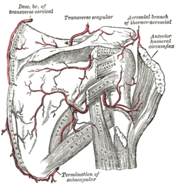
FIG. 524– The scapular and circumflex arteries. (Picture From the Classic Gray's Anatomy) - The Scapular Circumflex Artery (a. circumflexa scapulæ; dorsalis scapulæ artery) is generally larger than the continuation of the subscapular. It curves around the axillary border of the scapula, traversing the space between the Subscapularis above, the Teres major below, and the long head of the Triceps laterally (Fig. 524); it enters the infraspinatous fossa under cover of the Teres minor, and anastomoses with the transverse scapular artery and the descending branch of the transverse cervical. In its course it gives off two branches: one (infrascapular) enters the subscapular fossa beneath the Subscapularis, which it supplies, anastomosing with the transverse scapular artery and the descending branch of the transverse cervical; the other is continued along the axillary border of the scapula, between the Teres major and minor, and at the dorsal surface of the inferior angle anastomoses with the descending branch of the transverse cervical. In addition to these, small branches are distributed to the back part of the Deltoideus and the long head of the Triceps brachii, anastomosing with an ascending branch of the a. profunda brachii.
- 5. The posterior humeral circumflex artery (a. circumflexa humeri posterior; posterior circumflex artery) (Fig. 524) arises from the axillary artery at the lower border of the Subscapularis, and runs backward with the axillary nerve through the quadrangular space bounded by the Subscapularis and Teres minor above, the Teres major below, the long head of the Triceps brachii medially, and the surgical neck of the humerus laterally. It winds around the neck of the humerus and is distributed to the Deltoideus and shoulder-joint, anastomosing with the anterior humeral circumflex and profunda brachii.
- 6. The anterior humeral circumflex artery (a. circumflexa humeri anterior; anterior circumflex artery) (Fig. 524), considerably smaller than the posterior, arises nearly opposite it, from the lateral side of the axillary artery. It runs horizontally, beneath the Coracobrachialis and short head of the Biceps brachii, in front of the neck of the humerus. On reaching the intertubercular sulcus, it gives off a branch which ascends in the sulcus to supply the head of the humerus and the shoulder-joint. The trunk of the vessel is then continued onward beneath the long head of the Biceps brachii and the Deltoideus, and anastomoses with the posterior humeral circumflex artery.
- Peculiarities—The branches of the axillary artery vary considerably in different subjects. Occasionally the subscapular, humeral circumflex, and profunda arteries arise from a common trunk, and when this occurs the branches of the brachial plexus surround this trunk instead of the main vessel. Sometimes the axillary artery divides into the radial and ulnar arteries, and occasionally it gives origin to the volar interosseous artery of the forearm.

Gray's Anatomy[edit]
- Gray's Anatomy Contents
- Gray's Anatomy Subject Index
- About Classic Gray's Anatomy
- Glossary of anatomy terms
Anatomy atlases (external)[edit]
[1] - Anatomy Atlases
| Human systems and organs | ||||||||||||||
|---|---|---|---|---|---|---|---|---|---|---|---|---|---|---|
|

Medical Disclaimer: WikiMD is for informational purposes only and is not a substitute for professional medical advice. Content may be inaccurate or outdated and should not be used for diagnosis or treatment. Always consult your healthcare provider for medical decisions. Verify information with trusted sources such as CDC.gov and NIH.gov. By using this site, you agree that WikiMD is not liable for any outcomes related to its content. See full disclaimer.
Credits:Most images are courtesy of Wikimedia commons, and templates, categories Wikipedia, licensed under CC BY SA or similar.
Translate page: - East Asian
中文,
日本,
한국어,
South Asian
हिन्दी,
தமிழ்,
తెలుగు,
Urdu,
ಕನ್ನಡ,
Southeast Asian
Indonesian,
Vietnamese,
Thai,
မြန်မာဘာသာ,
বাংলা
European
español,
Deutsch,
français,
Greek,
português do Brasil,
polski,
română,
русский,
Nederlands,
norsk,
svenska,
suomi,
Italian
Middle Eastern & African
عربى,
Turkish,
Persian,
Hebrew,
Afrikaans,
isiZulu,
Kiswahili,
Other
Bulgarian,
Hungarian,
Czech,
Swedish,
മലയാളം,
मराठी,
ਪੰਜਾਬੀ,
ગુજરાતી,
Portuguese,
Ukrainian
