The Thyroid Gland
Anatomy > Gray's Anatomy of the Human Body > XI. Splanchnology > 4. The Ductless Glands
Henry Gray (1821–1865). Anatomy of the Human Body. 1918.
4. The Ductless Glands
There are certain organs which are very similar to secreting glands, but differ from them in one essential particular, viz., they do not possess any ducts by which their secretion is discharged.
These organs are known as ductless glands They are capable of internal secretion—that is to say, of forming, from materials brought to them in the blood, substances which have a certain influence upon the nutritive and other changes going on in the body. This secretion is carried into the blood stream, either directly by the veins or indirectly through the medium of the lymphatics.
These glands include the thyroid the parathyroids and the thymus the pituitary body and the pineal body the chromaphil and cortical systems to which belong the suprarenals the paraganglia and aortic glands the glomus caroticum and perhaps the glomus coccygeum The spleen is usually included in this list and sometimes the lymph and hemolymph nodes described with the lymphatic system. Other glands as the liver, pancreas and sexual glands give off internal secretions, as do the gastric and intestinal mucous membranes.
a. The Thyroid Gland (Glandula Thyreiodea; Thyroid Body) (Fig. 1174)—The thyroid gland is a highly vascular organ, situated at the front and sides of the neck; it consists of right and left lobes connected across the middle line by a narrow portion, the isthmus Its weight is somewhat variable, but is usually about 30 grams. It is slightly heavier in the female, in whom it becomes enlarged during menstruation and pregnancy.
The lobes (lobuli gl. thyreoideæ) are conical in shape, the apex of each being directed upward and lateralward as far as the junction of the middle with the lower third of the thyroid cartilage; the base looks downward, and is on a level with the fifth or sixth tracheal ring. Each lobe is about 5 cm. long; its greatest width is about 3 cm., and its thickness about 2 cm.
The lateral or superficial surface is convex, and covered by the skin, the superficial and deep fasciæ, the Sternocleidomastoideus, the superior belly of the Omohyoideus, the Sternohyoideus and Sternothyreoideus, and beneath the last muscle by the pretracheal layer of the deep fascia, which forms a capsule for the gland.
The deep or medial surface is moulded over the underlying structures, viz., the thyroid and cricoid cartilages, the trachea, the Constrictor pharyngis inferior and posterior part of the Cricothyreoideus, the esophagus (particularly on the left side of the neck), the superior and inferior thyroid arteries, and the recurrent nerves. The anterior border is thin, and inclines obliquely from above downward toward the middle line of the neck, while the posterior border is thick and overlaps the common carotid artery, and, as a rule, the parathyroids.
The isthmus (isthmus gl. thyreoidea) connects together the lower thirds of the lobes; it measures about 1.25 cm. in breadth, and the same in depth, and usually covers the second and third rings of the trachea. Its situation and size present, however, many variations. In the middle line of the neck it is covered by the skin and fascia, and close to the middle line, on either side, by the Sternothyreoideus. Across its upper border runs an anastomotic branch uniting the two superior thyroid arteries; at its lower border are the inferior thyroid veins. Sometimes the isthmus is altogether wanting.
A third lobe, of conical shape, called the pyramidal lobe frequently arises from the upper part of the isthmus, or from the adjacent portion of either lobe, but most commonly the left, and ascends as far as the hyoid bone. It is occasionally quite detached, or may be divided into two or more parts.
A fibrous or muscular band is sometimes found attached, above, to the body of the hyoid bone, and below to the isthmus of the gland, or its pyramidal lobe. When muscular, it is termed the Levator glandulæ thyreoideæ
Small detached portions of thyroid tissue are sometimes found in the vicinity of the lateral lobes or above the isthmus; they are called accessory thyroid glands (glandulæ thyreoideæ accessoriæ).
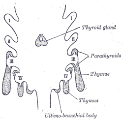
IV Branchial pouches. (Picture From the Classic Gray's Anatomy)
Development—The thyroid gland is developed from a median diverticulum (Fig. 1175), which appears about the fourth week on the summit of the tuberculum impar, but later is found in the furrow immediately behind the tuberculum (Fig. 979). It grows downward and backward as a tubular duct, which bifurcates and subsequently subdivides into a series of cellular cords, from which the isthmus and lateral lobes of the thyroid gland are developed.
The ultimo-branchial bodies from the fifth pharyngeal pouches are enveloped by the lateral lobes of the thyroid gland; they undergo atrophy and do not form true thyroid tissue. The connection of the diverticulum with the pharynx is termed the thyroglossal duct its continuity is subsequently interrupted, and it undergoes degeneration, its upper end being represented by the foramen cecum of the tongue, and its lower by the pyramidal lobe of the thyroid gland.
Structure—The thyroid gland is invested by a thin capsule of connective tissue, which projects into its substance and imperfectly divides it into masses of irregular form and size. When the organ is cut into, it is of a brownish-red color, and is seen to be made up of a number of closed vesicles, containing a yellow glairy fluid, and separated from each other by intermediate connective tissue (Fig. 1176).
The vesicles of the thyroid of the adult animal are generally closed spherical sacs; but in some young animals, e. g young dogs, the vesicles are more or less tubular and branched. This appearance is supposed to be due to the mode of growth of the gland, and merely indicates that an increase in the number of vesicles is taking place. Each vesicle is lined by a single layer of cubical epithelium. There does not appear to be a basement membrane, so that the epithelial cells are in direct contact with the connective-tissue reticulum which supports the acini. The vesicles are of various sizes and shapes, and contain as a normal product a viscid, homogeneous, semifluid, slightly yellowish, colloid material; red corpuscles are found in it in various stages of disintegration and decolorization, the yellow tinge being probably due to the hemoglobin, which is thus set free from the colored corpuscles.
The colloid material contains an iodine compound, iodothyrin and is readily stained by eosin. According to Bensley 180 the thyroid gland prepares and secretes into the vascular channels a substance, formed under normal conditions in the outer pole of the cell and excreted from it directly without passing by the indirect route through the follicular cavity. In addition to this direct mode of secretion there is an indirect mode which consists in the condensation of the secretion into the form of droplets, having high content of solids, and the extension of these droplets into the follicular cavity. These droplets are formed in the same zone of the cell as that in which the primary or direct secretion is formed.
This internal secretion of the thyroid is supposed to contain a specific hormone which acts as a chemical stimulus to other tissues, increasing their metabolism.
- Vessels and Nerves—The arteries supplying the thyroid gland are the superior and inferior thyroids and sometimes an additional branch (thyroidea ima) from the innominate artery or the arch of the aorta, which ascends upon the front of the trachea.
- The arteries are remarkable for their large size and frequent anastomoses. The veins form a plexus on the surface of the gland and on the front of the trachea; from this plexus the superior, middle, and inferior thyroid veins arise; the superior and middle end in the internal jugular, the inferior in the innominate vein.
- The capillary blood vessels form a dense plexus in the connective tissue around the vesicles, between the epithelium of the vesicles and the endothelium of the lymphatics, which surround a greater or smaller part of the circumference of the vesicle. The lymphatic vessels run in the interlobular connective tissue, not uncommonly surrounding the arteries which they accompany, and communicate with a net-work in the capsule of the gland; they may contain colloid material. They end in the thoracic and right lymphatic trunks. The nerves are derived from the middle and inferior cervical ganglia of the sympathetic.
Gray's Anatomy[edit source]
- Gray's Anatomy Contents
- Gray's Anatomy Subject Index
- About Classic Gray's Anatomy
- Glossary of anatomy terms
Anatomy atlases (external)[edit source]
[1] - Anatomy Atlases
| This article is a medical stub. You can help WikiMD by expanding it! | |
|---|---|
| Human systems and organs | ||||||||||||||
|---|---|---|---|---|---|---|---|---|---|---|---|---|---|---|
|
Search WikiMD
Ad.Tired of being Overweight? Try W8MD's NYC physician weight loss.
Semaglutide (Ozempic / Wegovy and Tirzepatide (Mounjaro / Zepbound) available. Call 718 946 5500.
Advertise on WikiMD
|
WikiMD's Wellness Encyclopedia |
| Let Food Be Thy Medicine Medicine Thy Food - Hippocrates |
Translate this page: - East Asian
中文,
日本,
한국어,
South Asian
हिन्दी,
தமிழ்,
తెలుగు,
Urdu,
ಕನ್ನಡ,
Southeast Asian
Indonesian,
Vietnamese,
Thai,
မြန်မာဘာသာ,
বাংলা
European
español,
Deutsch,
français,
Greek,
português do Brasil,
polski,
română,
русский,
Nederlands,
norsk,
svenska,
suomi,
Italian
Middle Eastern & African
عربى,
Turkish,
Persian,
Hebrew,
Afrikaans,
isiZulu,
Kiswahili,
Other
Bulgarian,
Hungarian,
Czech,
Swedish,
മലയാളം,
मराठी,
ਪੰਜਾਬੀ,
ગુજરાતી,
Portuguese,
Ukrainian
Medical Disclaimer: WikiMD is not a substitute for professional medical advice. The information on WikiMD is provided as an information resource only, may be incorrect, outdated or misleading, and is not to be used or relied on for any diagnostic or treatment purposes. Please consult your health care provider before making any healthcare decisions or for guidance about a specific medical condition. WikiMD expressly disclaims responsibility, and shall have no liability, for any damages, loss, injury, or liability whatsoever suffered as a result of your reliance on the information contained in this site. By visiting this site you agree to the foregoing terms and conditions, which may from time to time be changed or supplemented by WikiMD. If you do not agree to the foregoing terms and conditions, you should not enter or use this site. See full disclaimer.
Credits:Most images are courtesy of Wikimedia commons, and templates, categories Wikipedia, licensed under CC BY SA or similar.
Contributors: Prab R. Tumpati, MD


