Vas deferens
A duct in the male reproductive system


The vas deferens, also known as the ductus deferens, is a vital component of the male reproductive system. It is a muscular tube that transports sperm from the epididymis in anticipation of ejaculation.
Anatomy[edit]
The vas deferens is approximately 30 centimeters long and is a continuation of the epididymis. It ascends from the scrotum, passes through the inguinal canal, and enters the pelvic cavity. The vas deferens then travels posteriorly to the urinary bladder, where it joins with the duct of the seminal vesicle to form the ejaculatory duct.
Structure[edit]
The wall of the vas deferens is composed of three layers: an inner mucosa, a middle muscularis, and an outer adventitia. The muscularis layer is particularly thick, consisting of longitudinal and circular smooth muscle fibers, which facilitate the propulsion of sperm during ejaculation.
Blood Supply[edit]
The vas deferens receives its blood supply from the artery of the vas deferens, a branch of the superior vesical artery. Venous drainage is via the pampiniform plexus, which eventually drains into the testicular vein.
Function[edit]
The primary function of the vas deferens is to transport sperm from the epididymis to the ejaculatory duct. During ejaculation, the muscular walls of the vas deferens contract, propelling sperm forward. This process is facilitated by the presence of peristaltic waves.
Clinical Significance[edit]
The vas deferens is a key structure in vasectomy, a surgical procedure for male contraception. During a vasectomy, the vas deferens is cut and sealed to prevent sperm from entering the ejaculatory duct, thereby preventing fertilization.
Gallery[edit]
-
 The male reproductive organs.
 -
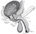
 The vas deferens and surrounding structures.
 -
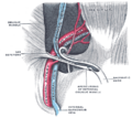
 The vas deferens in relation to the bladder.
 -
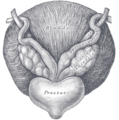
 The vas deferens and seminal vesicle.
 -
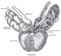
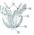 Illustration of the prostate and surrounding structures.
 -
Close-up of the vas deferens.
-
Testis and associated structures.
-
Histological slide of the vas deferens.


Related pages[edit]
References[edit]
- Moore, K. L., Dalley, A. F., & Agur, A. M. R. (2013). Clinically Oriented Anatomy. Lippincott Williams & Wilkins.
- Netter, F. H. (2014). Atlas of Human Anatomy. Elsevier.

Medical Disclaimer: WikiMD is for informational purposes only and is not a substitute for professional medical advice. Content may be inaccurate or outdated and should not be used for diagnosis or treatment. Always consult your healthcare provider for medical decisions. Verify information with trusted sources such as CDC.gov and NIH.gov. By using this site, you agree that WikiMD is not liable for any outcomes related to its content. See full disclaimer.
Credits:Most images are courtesy of Wikimedia commons, and templates, categories Wikipedia, licensed under CC BY SA or similar.
Translate page: - East Asian
中文,
日本,
한국어,
South Asian
हिन्दी,
தமிழ்,
తెలుగు,
Urdu,
ಕನ್ನಡ,
Southeast Asian
Indonesian,
Vietnamese,
Thai,
မြန်မာဘာသာ,
বাংলা
European
español,
Deutsch,
français,
Greek,
português do Brasil,
polski,
română,
русский,
Nederlands,
norsk,
svenska,
suomi,
Italian
Middle Eastern & African
عربى,
Turkish,
Persian,
Hebrew,
Afrikaans,
isiZulu,
Kiswahili,
Other
Bulgarian,
Hungarian,
Czech,
Swedish,
മലയാളം,
मराठी,
ਪੰਜਾਬੀ,
ગુજરાતી,
Portuguese,
Ukrainian
