The Anterior Tibial Artery
Anatomy > Gray's Anatomy of the Human Body > VI. The Arteries > 6d. The Anterior Tibial Artery
Henry Gray (1821–1865). Anatomy of the Human Body. 1918.
The Anterior Tibial Artery[edit]
(A. Tibialis Anterior)
The anterior tibial artery (Fig. 553) commences at the bifurcation of the popliteal, at the lower border of the Popliteus, passes forward between the two heads of the Tibialis posterior, and through the aperture above the upper border of the interosseous membrane, to the deep part of the front of the leg: it here lies close to the medial side of the neck of the fibula.
It then descends on the anterior surface of the interosseous membrane, gradually approaching the tibia; at the lower part of the leg it lies on this bone, and then on the front of the ankle-joint, where it is more superficial, and becomes the dorsalis pedis
Relations[edit]
In the upper two-thirds of its extent, the anterior tibial artery rests upon the interosseous membrane; in the lower third, upon the front of the tibia, and the anterior ligament of the ankle-joint.
In the upper third of its course, it lies between the Tibialis anterior and Extensor digitorum longus; in the middle third between the Tibialis anterior and Extensor hallucis longus. At the ankle it is crossed from the lateral to the medial side by the tendon of the Extensor hallucis longus, and lies between it and the first tendon of the Extensor digitorum longus.
It is covered in the upper two-thirds of its course, by the muscles which lie on either side of it, and by the deep fascia; in the lower third, by the integument and fascia, and the transverse and cruciate crural ligaments.
The anterior tibial artery is accompanied by a pair of venae comitantes which lie one on either side of the artery; the deep peroneal nerve, coursing around the lateral side of the neck of the fibula, comes into relation with the lateral side of the artery shortly after it has reached the front of the leg; about the middle of the leg the nerve is in front of the artery; at the lower part it is generally again on the lateral side.
Peculiarities in Size[edit]
This vessel may be diminished in size, may be deficient to a greater or less extent, or may be entirely wanting, its place being supplied by perforating branches from the posterior tibial, or by the perforating branch of the peroneal artery.
Course[edit]
The artery occasionally deviates toward the fibular side of the leg, regaining its usual position at the front of the ankle. In rare instances the vessel has been found to approach the surface in the middle of the leg, being covered merely by the integument and fascia below that point.
Branches[edit]
The branches of the anterior tibial artery are:
Anterior Tibial Recurrent.
Anterior Lateral Malleolar.
The posterior tibial recurrent artery[edit]
The posterior tibial recurrent artery (a. recurrens tibialis posterior) an inconstant branch, is given off from the anterior tibial before that vessel passes through the interosseous space. It ascends in front of the Popliteus, which it supplies, and anastomoses with the inferior genicular branches of the popliteal artery, giving an offset to the tibiofibular joint.
Fibular artery[edit]
The fibular artery is sometimes derived from the anterior tibial, sometimes from the posterior tibial. It passes lateralward, around the neck of the fibula, through the Soleus, which it supplies, and ends in the substance of the Peroneus longus.
The anterior tibial recurrent artery[edit]
The anterior tibial recurrent artery (a. recurrens tibialis anterior) arises from the anterior tibial, as soon as that vessel has passed through the interosseous space; it ascends in the Tibialis anterior, ramifies on the front and sides of the knee-joint, and assists in the formation of the patellar plexus by anastomosing with the genicular branches of the popliteal, and with the highest genicular artery.
The muscular branches[edit]
The muscular branches (rami musculares) are numerous; they are distributed to the muscles which lie on either side of the vessel, some piercing the deep fascia to supply the integument, others passing through the interosseous membrane, and anastomosing with branches of the posterior tibial and peroneal arteries.
The anterior medial malleolar artery[edit]
The anterior medial malleolar artery (a. malleolaris anterior medialis; internal malleolar artery) arises about 5 cm. above the ankle-joint and passes behind the tendons of the Extensor hallucis longus and Tibialis anterior, to the medial side of the ankle, upon which it ramifies, anastomosing with branches of the posterior tibial and medial plantar arteries and with the medial calcaneal from the posterior tibial.
The anterior lateral malleolar artery[edit]
The anterior lateral malleolar artery (a. malleolaris anterior lateralis; external malleolar artery) passes beneath the tendons of the Extensor digitorum longus and Peronaeus tertius and supplies the lateral side of the ankle, anastomosing with the perforating branch of the peroneal artery, and with ascending twigs from the lateral tarsal artery.
The arteries around the ankle-joint anastomose freely with one another and form net-works below the corresponding malleoli.
The medial malleolar net-work is formed by the anterior medial malleolar branch of the anterior tibial, the medial tarsal branches of the dorsalis pedis, the posterior medial malleolar and medial calcaneal branches of the posterior tibial and branches from the medial plantar artery.
The lateral malleolar net-work is formed by the anterior lateral malleolar branch of the anterior tibial, the lateral tarsal branch of the dorsalis pedis, the perforating and the lateral calcaneal branches of the peroneal, and twigs from the lateral plantar artery.

Clinical importance[edit]
As the artery passes medial to the fibular neck it becomes vulnerable to damage during a tibial osteotomy.
Additional images[edit]
-
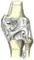 Right knee-joint. Posterior view.
 -
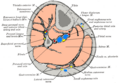 Cross-section through middle of leg.
 -
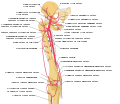 Schema of the arteries of the thigh. Anterior tibial artery is labeled at the bottom.
 -
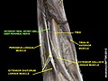 Anterior tibial artery
 -
 Anterior tibial artery
 -
 Anterior tibial artery


External links[edit]
- Gray's s157 - The Arteries of the Lower Extremity
- Gray's s95 - Ankle joint
- Anatomy figure: 12:04-15 at Human Anatomy Online, SUNY Downstate Medical Center - "Arteries of the lower extremity shown in association with major landmarks."
| Arteries of the human leg | ||||||||||||||||||||||||||||
|---|---|---|---|---|---|---|---|---|---|---|---|---|---|---|---|---|---|---|---|---|---|---|---|---|---|---|---|---|
|
Gray's Anatomy[edit]
- Gray's Anatomy Contents
- Gray's Anatomy Subject Index
- About Classic Gray's Anatomy
- Glossary of anatomy terms
Anatomy atlases (external)[edit]
[1] - Anatomy Atlases
| Human systems and organs | ||||||||||||||
|---|---|---|---|---|---|---|---|---|---|---|---|---|---|---|
|

Medical Disclaimer: WikiMD is for informational purposes only and is not a substitute for professional medical advice. Content may be inaccurate or outdated and should not be used for diagnosis or treatment. Always consult your healthcare provider for medical decisions. Verify information with trusted sources such as CDC.gov and NIH.gov. By using this site, you agree that WikiMD is not liable for any outcomes related to its content. See full disclaimer.
Credits:Most images are courtesy of Wikimedia commons, and templates, categories Wikipedia, licensed under CC BY SA or similar.
Translate page: - East Asian
中文,
日本,
한국어,
South Asian
हिन्दी,
தமிழ்,
తెలుగు,
Urdu,
ಕನ್ನಡ,
Southeast Asian
Indonesian,
Vietnamese,
Thai,
မြန်မာဘာသာ,
বাংলা
European
español,
Deutsch,
français,
Greek,
português do Brasil,
polski,
română,
русский,
Nederlands,
norsk,
svenska,
suomi,
Italian
Middle Eastern & African
عربى,
Turkish,
Persian,
Hebrew,
Afrikaans,
isiZulu,
Kiswahili,
Other
Bulgarian,
Hungarian,
Czech,
Swedish,
മലയാളം,
मराठी,
ਪੰਜਾਬੀ,
ગુજરાતી,
Portuguese,
Ukrainian
