The Tarsus
Anatomy > Gray's Anatomy of the Human Body > II. [Osteology]] > 6d. The Foot. 1. The Tarsus
Henry Gray (1821–1865). Anatomy of the Human Body. 1918.
The Tarsus[edit | edit source]
(ossa tarsi)
The skeleton of the foot (Figs. 268 and 269) consists of three parts: the tarsus, metatarsus and phalanges
The tarsal bones are seven in number, viz., the calcaneus, talus, cuboid, navicular and the first, second and third cuneiforms
The Calcaneus (os calcis) (Figs. 264 to 267)[edit | edit source]
The calcaneus is the largest of the tarsal bones. It is situated at the lower and back part of the foot, serving to transmit the weight of the body to the ground, and forming a strong lever for the muscles of the calf. It is irregularly cuboidal in form, having its long axis directed forward and lateralward; it presents for examination six surfaces.
FIG. 266– Left calcaneus, lateral surface. (Picture From the Classic Gray's Anatomy)
FIG. 267– Left calcaneus, medial surface. (Picture From the Classic Gray's Anatomy)
Surfaces[edit | edit source]
The superior surface extends behind on to that part of the bone which projects backward to form the heel. This varies in length in different individuals, is convex from side to side, concave from before backward, and supports a mass of fat placed in front of the tendo calcaneus.
In front of this area is a large usually somewhat oval-shaped facet, the posterior articular surface which looks upward and forward; it is convex from behind forward, and articulates with the posterior calcaneal facet on the under surface of the talus.
It is bounded anteriorly by a deep depression which is continued backward and medialward in the form of a groove, the calcaneal sulcus In the articulated foot this sulcus lies below a similar one on the under surface of the talus, and the two form a canal (sinus tarsi) for the lodgement of the interosseous talocalcaneal ligament.
In front and to the medial side of this groove is an elongated facet, concave from behind forward, and with its long axis directed forward and lateralward. This facet is frequently divided into two by a notch: of the two, the posterior, and larger is termed the middle articular surface it is supported on a projecting process of bone, the sustentaculum tali and articulates with the middle calcaneal facet on the under surface of the talus; the anterior articular surface is placed on the anterior part of the body, and articulates with the anterior calcaneal facet on the talus. The upper surface, anterior and lateral to the facets, is rough for the attachment of ligaments and for the origin of the Extensor digitorum brevis. [[File:Gray268.gif
FIG. 268– Bones of the right foot. Dorsal surface. (Picture From the Classic Gray's Anatomy)
FIG. 269– Bones of the right foot. Plantar surface. (Picture From the Classic Gray's Anatomy)
The inferior or plantar surface is uneven, wider behind than in front, and convex from side to side; it is bounded posteriorly by a transverse elevation, the calcaneal tuberosity which is depressed in the middle and prolonged at either end into a process; the lateral process small, prominent, and rounded, gives origin to part of the Abductor digiti quinti; the medial process broader and larger, gives attachment, by its prominent medial margin, to the Abductor hallucis, and in front to the Flexor digitorum brevis and the plantar aponeurosis; the depression between the processes gives origin to the Abductor digiti quinti. The rough surface in front of the processes gives attachment to the long plantar ligament, and to the lateral head of the Quadratus plantae while to a prominent tubercle nearer the anterior part of this surface, as well as to a transverse groove in front of the tubercle, is attached the plantar calcaneocuboid ligament.
The Lateral surface is broad behind and narrow in front, flat and almost subcutaneous; near its center is a tubercle, for the attachment of the calcaneofibular ligament. At its upper and anterior part, this surface gives attachment to the lateral talocalcaneal ligament; and in front of the tubercle it presents a narrow surface marked by two oblique grooves. The grooves are separated by an elevated ridge, or tubercle, the trochlear process (peroneal tubercle), which varies much in size in different bones.
The superior groove transmits the tendon of the Peronaeus brevis; the inferior groove that of the Peronaeus longus.
The Medial surface is deeply concave; it is directed obliquely downward and forward, and serves for the transmission of the plantar vessels and nerves into the sole of the foot; it affords origin to part of the Quadratus plantae. At its upper and forepart is a horizontal eminence, the sustentaculum tali which gives attachment to a slip of the tendon of the Tibialis posterior. This eminence is concave above, and articulates with the middle calcaneal articular surface of the talus; below, it is grooved for the tendon of the Flexor hallucis longus; its anterior margin gives attachment to the plantar calcaneonavicular ligament, and its medial, to a part of the deltoid ligament of the ankle-joint.
The anterior or cuboid articular surface is of a somewhat triangular form. It is concave from above downward and lateralward, and convex in a direction at right angles to this. Its medial border gives attachment to the plantar calcaneonavicular ligament.
The posterior surface is prominent, convex, wider below than above, and divisible into three areas. The lowest of these is rough, and covered by the fatty and fibrous tissue of the heel; the middle, also rough, gives insertion to the tendo calcaneus and Plantaris; while the highest is smooth, and is covered by a bursa which intervenes between it and the tendo calcaneus.
Articulations[edit | edit source]
The calcaneus articulates with two bones: the talus and cuboid.
The Talus (astragalus; ankle bone) (Figs. 270 to 273)[edit | edit source]
The talus is the second largest of the tarsal bones. It occupies the middle and upper part of the tarsus, supporting the tibia above, resting upon the calcaneus below, articulating on either side with the malleoli, and in front with the navicular. It consists of a body a neck and a head
The Body (corpus tali)[edit | edit source]
The superior surface of the body presents, behind, a smooth trochlear surface, the trochlea for articulation with the tibia. The trochlea is broader in front than behind, convex from before backward, slightly concave from side to side: in front it is continuous with the upper surface of the neck of the bone.
FIG. 270– Left talus, from above. (Picture From the Classic Gray's Anatomy)
FIG. 271– Left talus, from below. (Picture From the Classic Gray's Anatomy)
The inferior surface presents two articular areas, the posterior and middle calcaneal surfaces, separated from one another by a deep groove, the sulcus tali The groove runs obliquely forward and lateralward, becoming gradually broader and deeper in front: in the articulated foot it lies above a similar groove upon the upper surface of the calcaneus, and forms, with it, a canal (sinus tarsi) filled up in the fresh state by the interosseous talocalcaneal ligament.
The posterior calcaneal articular surface is large and of an oval or oblong form. It articulates with the corresponding facet on the upper surface of the calcaneus, 63 and is deeply concave in the direction of its long axis which runs forward and lateralward at an angle of about 45° with the median plane of the body.
The middle calcaneal articular surface is small, oval in form and slightly convex; it articulates with the upper surface of the sustentaculum tali of the calcaneus.
The medial surface presents at its upper part a pear-shaped articular facet for the medial malleolus, continuous above with the trochlea; below the articular surface is a rough depression for the attachment of the deep portion of the deltoid ligament of the ankle-joint.
FIG. 272– Left talus, medial surface. (Picture From the Classic Gray's Anatomy)
FIG. 273– Left talus, lateral surface. (Picture From the Classic Gray's Anatomy)
The lateral surface carries a large triangular facet, concave from above downward, for articulation with the lateral malleolus; its anterior half is continuous above with the trochlea; and in front of it is a rough depression for the attachment of the anterior talofibular ligament. Between the posterior half of the lateral border of the trochlea and the posterior part of the base of the fibular articular surface is a triangular facet (Fawcett 64) which comes into contact with the transverse inferior tibiofibular ligament during flexion of the ankle-joint; below the base of this facet is a groove which affords attachment to the posterior talofibular ligament.
The posterior surface is narrow, and traversed by a groove running obliquely downward and medialward, and transmitting the tendon of the Flexor hallucis longus. Lateral to the groove is a prominent tubercle, the posterior process to which the posterior talofibular ligament is attached; this process is sometimes separated from the rest of the talus, and is then known as the os trigonum Medial to the groove is a second smaller tubercle.
The Neck (collum tali)[edit | edit source]
The neck is directed forward and medialward, and comprises the constricted portion of the bone between the body and the oval head. Its upper and medial surfaces are rough, for the attachment of ligaments; its lateral surface is concave and is continuous below with the deep groove for the interosseous talocalcaneal ligament. 17
The Head (caput tali)[edit | edit source]
The head looks forward and medialward; its anterior articular or navicular surface is large, oval, and convex. Its inferior surface has two facets, which are best seen in the fresh condition. The medial, situated in front of the middle calcaneal facet, is convex, triangular, or semi-oval in shape, and rests on the plantar calcaneonavicular ligament; the lateral, named the anterior calcaneal articular surface is somewhat flattened, and articulates with the facet on the upper surface of the anterior part of the calcaneus.
Articulations[edit | edit source]
The talus articulates with four bones: tibia, fibula, calcaneus, and navicular.
The Cuboid Bone (os cuboideum) (Figs. 274, 275)[edit | edit source]
The cuboid bone is placed on the lateral side of the foot, in front of the calcaneus, and behind the fourth and fifth metatarsal bones. It is of a pyramidal shape, its base being directed medialward.
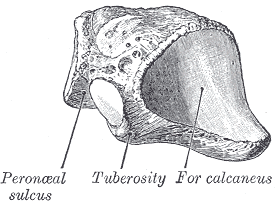
FIG. 274– The left cuboid. Antero-medial view. (Picture From the Classic Gray's Anatomy)
FIG. 275– The left cuboid. Postero-lateral view. (Picture From the Classic Gray's Anatomy)
Surfaces[edit | edit source]
The dorsal surface directed upward and lateralward, is rough, for the attachment of ligaments. The plantar surface presents in front a deep groove, the peroneal sulcus which runs obliquely forward and medialward; it lodges the tendon of the Peronaeus longus, and is bounded behind by a prominent ridge, to which the long plantar ligament is attached. The ridge ends laterally in an eminence, the tuberosity the surface of which presents an oval facet; on this facet glides the sesamoid bone or cartilage frequently found in the tendon of the Peronaeus longus. The surface of bone behind the groove is rough, for the attachment of the plantar calcaneocuboid ligament, a few fibers of the Flexor hallucis brevis, and a fasciculus from the tendon of the Tibialis posterior. The lateral surface presents a deep notch formed by the commencement of the peroneal sulcus. The posterior surface is smooth, triangular, and concavo-convex, for articulation with the anterior surface of the calcaneus; its infero-medial angle projects backward as a process which underlies and supports the anterior end of the calcaneus. The anterior surface of smaller size, but also irregularly triangular, is divided by a vertical ridge into two facets: the medial, quadrilateral in form, articulates with the fourth metatarsal; the lateral, larger and more triangular, articulates with the fifth. The medial surface is broad, irregularly quadrilateral, and presents at its middle and upper part a smooth oval facet, for articulation with the third cuneiform; and behind this (occasionally) a smaller facet, for articulation with the navicular; it is rough in the rest of its extent, for the attachment of strong interosseous ligaments.
Articulations[edit | edit source]
The cuboid articulates with four bones: the calcaneus, third cuneiform, and fourth and fifth metatarsals; occasionally with a fifth, the navicular.
[edit | edit source]
The navicular bone is situated at the medial side of the tarsus, between the talus behind and the cuneiform bones in front.
FIG. 276– The left navicular. Antero-lateral view. (Picture From the Classic Gray's Anatomy)
FIG. 277– The left navicular. Postero-medial view. (Picture From the Classic Gray's Anatomy)
Surfaces[edit | edit source]
The anterior surface is convex from side to side, and subdivided by two ridges into three facets, for articulation with the three cuneiform bones. The posterior surface is oval, concave, broader laterally than medially, and articulates with the rounded head of the talus. The dorsal surface is convex from side to side, and rough for the attachment of ligaments. The plantar surface is irregular, and also rough for the attachment of ligaments. The medial surface presents a rounded tuberosity the lower part of which gives attachment to part of the tendon of the Tibialis posterior. The lateral surface is rough and irregular for the attachment of ligaments, and occasionally presents a small facet for articulation with the cuboid bone.
Articulations[edit | edit source]
The navicular articulates with four bones: the talus and the three cuneiforms; occasionally with a fifth, the cuboid.
The First Cuneiform Bone (os cuneiform primum; internalcuneiform) (Figs. 278, 279)[edit | edit source]
The first cuneiform bone is the largest of the three cuneiforms. It is situated at the medial side of the foot, between the navicular behind and the base of the first metatarsal in front.
FIG. 278– The left first cuneiform. Antero-medial view. (Picture From the Classic Gray's Anatomy)
FIG. 279– The left first cuneiform. Postero-lateral view. (Picture From the Classic Gray's Anatomy)
Surfaces[edit | edit source]
The medial surface is subcutaneous, broad, and quadrilateral; at its anterior plantar angle is a smooth oval impression, into which part of the tendon of the Tibialis anterior is inserted; in the rest of its extent it is rough for the attachment of ligaments.
The lateral surface is concave, presenting, along its superior and posterior borders a narrow L-shaped surface, the vertical limb and posterior part of the horizontal limb of which articulate with the second cuneiform, while the anterior part of the horizontal limb articulates with the second metatarsal bone: the rest of this surface is rough for the attachment of ligaments and part of the tendon of the Peronaeus longus.
The anterior surface kidney-shaped and much larger than the posterior, articulates with the first metatarsal bone.
The posterior surface is triangular, concave, and articulates with the most medial and largest of the three facets on the anterior surface of the navicular.
The plantar surface is rough, and forms the base of the wedge; at its back part is a tuberosity for the insertion of part of the tendon of the Tibialis posterior. It also gives insertion in front to part of the tendon of the Tibialis anterior.
The dorsal surface is the narrow end of the wedge, and is directed upward and lateralward; it is rough for the attachment of ligaments.
Articulations[edit | edit source]
The first cuneiform articulates with four bones: the navicular, second cuneiform, and first and second metatarsals.
The Second Cuneiform Bone (os cuneiforme secundum; middle cuneiform) (Figs. 280, 281)[edit | edit source]
The second cuneiform bone, the smallest of the three, is of very regular wedge-like form, the thin end being directed downward. It is situated between the other two cuneiforms, and articulates with the navicular behind, and the second metatarsal in front.
Surfaces[edit | edit source]
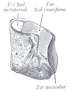
The anterior surface triangular in form, and narrower than the posterior, articulates with the base of the second metatarsal bone. The posterior surface also triangular, articulates with the intermediate facet on the anterior surface of the navicular.
The medial surface carries an L-shaped articular facet, running along the superior and posterior borders, for articulation with the first cuneiform, and is rough in the rest of its extent for the attachment of ligaments.
The lateral surface presents posteriorly a smooth facet for articulation with the third cuneiform bone.
The dorsal surface forms the base of the wedge; it is quadrilateral and rough for the attachment of ligaments.
The plantar surface sharp and tuberculated, is also rough for the attachment of ligaments, and for the insertion of a slip from the tendon of the Tibialis posterior.
FIG. 280– The left second cuneiform. Antero-medial view. (Picture From the Classic Gray's Anatomy)
FIG. 281– The left second cuneiform. Postero-lateral view. (Picture From the Classic Gray's Anatomy)
Articulations[edit | edit source]
The second cuneiform articulates with four bones: the navicular, first and third cuneiforms, and second metatarsal.
The Third Cuneiform Bone (os cuneiforme tertium; external cuneiform) (Figs. 282, 283)[edit | edit source]
The third cuneiform bone, intermediate in size between the two preceding, is wedge-shaped, the base being uppermost. It occupies the center of the front row of the tarsal bones, between the second cuneiform medially, the cuboid laterally, the navicular behind, and the third metatarsal in front.
Surfaces[edit | edit source]
The anterior surface triangular in form, articulates with the third metatarsal bone.
The [[posterior surface articulates with the lateral facet on the anterior surface of the navicular, and is rough below for the attachment of ligamentous fibers.
The medial surface presents an anterior and a posterior articular facet, separated by a rough depression: the anterior, sometimes divided, articulates with the lateral side of the base of the second metatarsal bone; the posterior skirts the posterior border, and articulates with the second cuneiform; the rough depression gives attachment to an interosseous ligament.
The lateral surface also presents two articular facets, separated by a rough non-articular area; the anterior facet, situated at the superior angle of the bone, is small and semi-oval in shape, and articulates with the medial side of the base of the fourth metatarsal bone; the posterior and larger one is triangular or oval, and articulates with the cuboid; the rough, non-articular area serves for the attachment of an interosseous ligament. The three facets for articulation with the three metatarsal bones are continuous with one another; those for articulation with the second cuneiform and navicular are also continuous, but that for articulation with the cuboid is usually separate.
The dorsal surface is of an oblong form, its postero-lateral angle being prolonged backward.
The plantar surface is a rounded margin, and serves for the attachment of part of the tendon of the Tibialis posterior, part of the Flexor hallucis brevis, and ligaments.
Articulations[edit | edit source]
The third cuneiform articulates with six bones: the navicular, second cuneiform, cuboid, and second, third, and fourth metatarsals.
FIG. 282– The left third cuneiform. Postero-medial view. (Picture From the Classic Gray's Anatomy)
FIG. 283– The third left cuneiform. Antero-lateral view. (Picture From the Classic Gray's Anatomy)
Note 63 Sewell (Journal of Anatomy and Physiology, vol. xxxviii) pointed out that in about 10 per cent. of bones a small triangular facet, continuous with the posterior calcaneal facet, is present at the junction of the lateral surface of the body with the posterior wall of the sulcus tali.
Note 64 Edinburgh Medical Journal, 1895.
External links[edit | edit source]
- Diagram, identifying bones
- xrayslowerlimb at The Anatomy Lesson by Wesley Norman (Georgetown University)
(xrayfootdorsal
)
| Bones of the human leg | ||||||||||||||||||||||||||
|---|---|---|---|---|---|---|---|---|---|---|---|---|---|---|---|---|---|---|---|---|---|---|---|---|---|---|
|
Gray's Anatomy[edit source]
- Gray's Anatomy Contents
- Gray's Anatomy Subject Index
- About Classic Gray's Anatomy
- Glossary of anatomy terms
Anatomy atlases (external)[edit source]
[1] - Anatomy Atlases
| This article is a medical stub. You can help WikiMD by expanding it! | |
|---|---|
| Human systems and organs | ||||||||||||||
|---|---|---|---|---|---|---|---|---|---|---|---|---|---|---|
|
Search WikiMD
Ad.Tired of being Overweight? Try W8MD's NYC physician weight loss.
Semaglutide (Ozempic / Wegovy and Tirzepatide (Mounjaro / Zepbound) available. Call 718 946 5500.
Advertise on WikiMD
|
WikiMD's Wellness Encyclopedia |
| Let Food Be Thy Medicine Medicine Thy Food - Hippocrates |
Translate this page: - East Asian
中文,
日本,
한국어,
South Asian
हिन्दी,
தமிழ்,
తెలుగు,
Urdu,
ಕನ್ನಡ,
Southeast Asian
Indonesian,
Vietnamese,
Thai,
မြန်မာဘာသာ,
বাংলা
European
español,
Deutsch,
français,
Greek,
português do Brasil,
polski,
română,
русский,
Nederlands,
norsk,
svenska,
suomi,
Italian
Middle Eastern & African
عربى,
Turkish,
Persian,
Hebrew,
Afrikaans,
isiZulu,
Kiswahili,
Other
Bulgarian,
Hungarian,
Czech,
Swedish,
മലയാളം,
मराठी,
ਪੰਜਾਬੀ,
ગુજરાતી,
Portuguese,
Ukrainian
Medical Disclaimer: WikiMD is not a substitute for professional medical advice. The information on WikiMD is provided as an information resource only, may be incorrect, outdated or misleading, and is not to be used or relied on for any diagnostic or treatment purposes. Please consult your health care provider before making any healthcare decisions or for guidance about a specific medical condition. WikiMD expressly disclaims responsibility, and shall have no liability, for any damages, loss, injury, or liability whatsoever suffered as a result of your reliance on the information contained in this site. By visiting this site you agree to the foregoing terms and conditions, which may from time to time be changed or supplemented by WikiMD. If you do not agree to the foregoing terms and conditions, you should not enter or use this site. See full disclaimer.
Credits:Most images are courtesy of Wikimedia commons, and templates, categories Wikipedia, licensed under CC BY SA or similar.
Contributors: Deepika vegiraju, Prab R. Tumpati, MD


