The Fibula
Anatomy > Gray's Anatomy of the Human Body > II. [Osteology]] > 6c. 6. The Fibula
Henry Gray (1821–1865). Anatomy of the Human Body. 1918.
The Fibula[edit]
(Calf Bone)
The fibula (Figs. 258, 259) is placed on the lateral side of the tibia, with which it is connected above and below. It is the smaller of the two bones, and, in proportion to its length, the most slender of all the long bones.
Its upper extremity is small, placed toward the back of the head of the tibia, below the level of the knee-joint, and excluded from the formation of this joint.
Its lower extremity inclines a little forward, so as to be on a plane anterior to that of the upper end; it projects below the tibia, and forms the lateral part of the ankle-joint. The bone has a body and two extremities
The Upper Extremity or Head (capitulum fibulœ; proximal extremity)[edit]
The upper extremity is of an irregular quadrate form, presenting above a flattened articular surface, directed upward, forward, and medialward, for articulation with a corresponding surface on the lateral condyle of the tibia. On the lateral side is a thick and rough prominence continued behind into a pointed eminence, the apex (styloid process), which projects upward from the posterior part of the head.
The prominence, at its upper and lateral part, gives attachment to the tendon of the Biceps femoris and to the fibular collateral ligament of the knee-joint, the ligament dividing the tendon into two parts. The remaining part of the circumference of the head is rough, for the attachment of muscles and ligaments.
It presents in front a tubercle for the origin of the upper and anterior fibers of the Peronaeus longus, and a surface for the attachment of the anterior ligament of the head; and behind, another tubercle, for the attachment of the posterior ligament of the head and the origin of the upper fibers of the Soleus.
The Body or Shaft (corpus fibulae)[edit]
The body presents four borders—the antero-lateral, the antero-medial, the postero-lateral, and the postero-medial; and four surfaces—anterior, posterior, medial, and lateral.
Borders[edit]
The antero-lateral border begins above in front of the head, runs vertically downward to a little below the middle of the bone, and then curving somewhat lateralward, bifurcates so as to embrace a triangular subcutaneous surface immediately above the lateral malleolus. This border gives attachment to an intermuscular septum, which separates the Extensor muscles on the anterior surface of the leg from the Peronaei longus and brevis on the lateral surface.
The antero-medial border or interosseous crest is situated close to the medial side of the preceding, and runs nearly parallel with it in the upper third of its extent, but diverges from it in the lower two-thirds. It begins above just beneath the head of the bone (sometimes it is quite indistinct for about 2.5 cm. below the head), and ends at the apex of a rough triangular surface immediately above the articular facet of the lateral malleolus. It serves for the attachment of the interosseous membrane, which separates the Extensor muscles in front from the Flexor muscles behind.


The postero-lateral border is prominent; it begins above at the apex, and ends below in the posterior border of the lateral malleolus. It is directed lateralward above, backward in the middle of its course, backward, and a little medialward below, and gives attachment to an aponeurosis which separates the Peronaei on the lateral surface from the Flexor muscles on the posterior surface.
The postero-medial border sometimes called the oblique line begins above at the medial side of the head, and ends by becoming continuous with the interosseous crest at the lower fourth of the bone. It is well-marked and prominent at the upper and middle parts of the bone. It gives attachment to an aponeurosis which separates the Tibialis posterior from the Soleus and Flexor hallucis longus.


Surfaces[edit]
The anterior surface is the interval between the antero-lateral and antero-medial borders. It is extremely narrow and flat in the upper third of its extent; broader and grooved longitudinally in its lower third; it serves for the origin of three muscles: the Extensor digitorum longus, Extensor hallucis longus, and Peronaeus tertius.
The posterior surface is the space included between the postero-lateral and the postero-medial borders; it is continuous below with the triangular area above the articular surface of the lateral malleolus; it is directed backward above, backward and medialward at its middle, directly medialward below. Its upper third is rough, for the origin of the Soleus; its lower part presents a triangular surface, connected to the tibia by a strong interosseous ligament; the intervening part of the surface is covered by the fibers of origin of the Flexor hallucis longus. Near the middle of this surface is the nutrient foramen, which is directed downward.
The medial surface is the interval included between the antero-medial and the postero-medial borders. It is grooved for the origin of the Tibialis posterior.
The lateral surface is the space between the antero-lateral and postero-lateral borders. It is broad, and often deeply grooved; it is directed lateralward in the upper two-thirds of its course, backward in the lower third, where it is continuous with the posterior border of the lateral malleolus. This surface gives origin to the Peronaei longus and brevis.
The Lower Extremity or Lateral Malleolus (malleolus lateralis; distal extremity; external malleolus)[edit]
The lower extremity is of a pyramidal form, and somewhat flattened from side to side; it descends to a lower level than the medial malleolus. The lateral surface is convex, subcutaneous, and continuous with the triangular, subcutaneous surface on the lateral side of the body.
The medial surface (Fig. 262) presents in front a smooth triangular surface, convex from above downward, which articulates with a corresponding surface on the lateral side of the talus. Behind and beneath the articular surface is a rough depression, which gives attachment to the posterior talofibular ligament.
The anterior border is thick and rough, and marked below by a depression for the attachment of the anterior talofibular ligament. The posterior border is broad and presents the shallow malleolar sulcus for the passage of the tendons of the Peronaei longus and brevis. The summit is rounded, and give attachment to the clacaneofibular ligament.
Ossification[edit]
The fibula is ossified from three centers (Fig. 263): one for the body, and one for either end. Ossification begins in the body about the eighth week of fetal life, and extends toward the extremities.
At birth the ends are cartilaginous. Ossification commences in the lower end in the second year, and in the upper about the fourth year. The lower epiphysis, the first to ossify, unites with the body about the twentieth year; the upper epiphysis joins about the twenty-fifth year.
Function[edit]
The fibula does not carry any significant load (weight) of the body. It extends past the lower end of the tibia and forms the outer part of the ankle providing stability to this joint. It has grooves for certain ligaments which gives them leverage and multiplies the muscle force. It provides attachment points for the following muscles:
 |
 |
| Muscle | Direction | Attachment[1] |
| Biceps femoris muscle | Insertion | Head of fibula |
| Extensor hallucis longus muscle | Origin | Medial side of fibula |
| Extensor digitorum longus muscle | Origin | Proximal part of the medial side of fibula |
| Fibularis tertius | Origin | Distal part of the medial side of fibula |
| Fibularis longus | Origin | Head and the lateral side of fibula |
| Fibularis brevis | Origin | Distal 2/3 of the lateral side of fibula |
| Soleus muscle | Origin | Proximal 1/3 of the posterior side of fibula |
| Tibialis posterior muscle | Origin | Lateral part of the posterior side of fibula |
| Flexor hallucis longus muscle | Origin | Posterior side of fibula |
Additional images[edit]
-
Position of fibula (shown in red)
-
3D image
-
Shape of fibula (right)
-
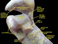 Ankle joint. Deep dissection.
 -
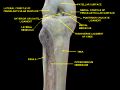 Knee and tibiofibular joint. Deep dissection. Anterior view.


External links[edit]
- Anatomy photo:17:st-1402 at the SUNY Downstate Medical Center
| Bones of the human leg | ||||||||||||||||||||||||||
|---|---|---|---|---|---|---|---|---|---|---|---|---|---|---|---|---|---|---|---|---|---|---|---|---|---|---|
|
Gray's Anatomy[edit]
- Gray's Anatomy Contents
- Gray's Anatomy Subject Index
- About Classic Gray's Anatomy
- Glossary of anatomy terms
Anatomy atlases (external)[edit]
[1] - Anatomy Atlases
| Human systems and organs | ||||||||||||||
|---|---|---|---|---|---|---|---|---|---|---|---|---|---|---|
|
- ↑ Finn, Bevægeapparatets anatomi, 12th edition, 2001, ISBN 978-87-628-0307-7, Pages: 364–367,

Medical Disclaimer: WikiMD is for informational purposes only and is not a substitute for professional medical advice. Content may be inaccurate or outdated and should not be used for diagnosis or treatment. Always consult your healthcare provider for medical decisions. Verify information with trusted sources such as CDC.gov and NIH.gov. By using this site, you agree that WikiMD is not liable for any outcomes related to its content. See full disclaimer.
Credits:Most images are courtesy of Wikimedia commons, and templates, categories Wikipedia, licensed under CC BY SA or similar.
Translate page: - East Asian
中文,
日本,
한국어,
South Asian
हिन्दी,
தமிழ்,
తెలుగు,
Urdu,
ಕನ್ನಡ,
Southeast Asian
Indonesian,
Vietnamese,
Thai,
မြန်မာဘာသာ,
বাংলা
European
español,
Deutsch,
français,
Greek,
português do Brasil,
polski,
română,
русский,
Nederlands,
norsk,
svenska,
suomi,
Italian
Middle Eastern & African
عربى,
Turkish,
Persian,
Hebrew,
Afrikaans,
isiZulu,
Kiswahili,
Other
Bulgarian,
Hungarian,
Czech,
Swedish,
മലയാളം,
मराठी,
ਪੰਜਾਬੀ,
ગુજરાતી,
Portuguese,
Ukrainian
