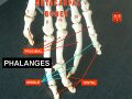
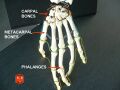
The Phalanges of the Hand
Anatomy > Gray's Anatomy of the Human Body > II. [Osteology]] > 6b. 3. The Phalanges of the Hand
Henry Gray (1821–1865). Anatomy of the Human Body. 1918.
The Phalanges of the Hand[edit]
(Phalanges Digitorum Manus)
- The phalanges are fourteen in number, three for each finger, and two for the thumb. Each consists of a body and two extremities.
- The body tapers from above downward, is convex posteriorly, concave in front from above downward, flat from side to side; its sides are marked by rough which give attachment to the fibrous sheaths of the Flexor tendons.
- The proximal extremities of the bones of the first row present oval, concave articular surfaces, broader from side to side than from before backward. The proximal extremity of each of the bones of the second and third rows presents a double concavity separated by a median ridge.
- The distal extremities are smaller than the proximal, and each ends in two condyles separated by a shallow groove; the articular surface extends farther on the volar than on the dorsal surface, a condition best marked in the bones of the first row.
- The ungual phalanges are convex on their dorsal and flat on their volar surfaces; they are recognized by their small size, and by a roughened, elevated surface of a horseshoe form on the volar surface of the distal extremity of each which serves to support the sensitive pulp of the finger.

Articulations[edit]
In the four fingers the phalanges of the first row articulate with those of the second row and with the metacarpals; the phalanges of the second row with those of the first and third rows, and the ungual phalanges with those of the second row. In the thumb, which has only two phalanges, the first phalanx articulates by its proximal extremity with the metacarpal bone and by its distal with the ungual phalanx.
Ossification of the Bones of the Hand[edit]
The carpal bones are each ossified from a single center, and ossification proceeds in the following order (Fig. 234): in the capitate and hamate, during the first year, the former preceding the latter; in the triangular, during the third year; in the lunate and greater multangular, during the fifth year, the former preceding the latter; in the navicular, during the sixth year; in the lesser multangular, during the eighth year; and in the pisiform, about the twelfth year Occasionally an additional bone, the os centrale is found on the back of the carpus, lying between the navicular, lesser multangular, and capitate. During the second month of fetal life it is represented by a small cartilaginous nodule, which usually fuses with the cartilaginous navicular. Sometimes the styloid process of the third metacarpal is detached and forms an additional ossicle.
The metacarpal bones are each ossified from two centers: one for the body and one for the distal extremity of each of the second, third, fourth, and fifth bones; one for the body and one for the base of the first metacarpal bone. 57 The first metacarpal bone is therefore ossified in the same manner as the phalanges, and this has led some anatomists to regard the thumb as being made up of three phalanges, and not of a metacarpal bone and two phalanges.
Ossification commences in the middle of the body about the eighth or ninth week of fetal life, the centers for the second and third metacarpals being the first, and that for the first metacarpal, the last, to appear; about the third year the distal extremities of the metacarpals of the fingers, and the base of the metacarpal of the thumb, begin to ossify; they unite with the bodies about the twentieth year.
The phalanges are each ossified from two centers: one for the body, and one for the proximal extremity. Ossification begins in the body, about the eighth week of fetal life. Ossification of the proximal extremity commences in the bones of the first row between the third and fourth years, and a year later in those of the second and third rows. The two centers become united in each row between the eighteenth and twentieth years. In the ungual phalanges the centers for the bodies appear at the distal extremities of the phalanges, instead of at the middle of the bodies, as in the other phalanges. Moreover, of all the bones of the hand, the ungual phalanges are the first to ossify.
Note 57 Allen Thomson demonstrated the fact that the first metacarpal bone is often developed from three centers: that is to say, there is a separate nucleus for the distal end, forming a distinct epiphysis visible at the age of seven or eight years. He also stated that there are traces of a proximal epiphysis in the second metacarpal bone, Journal of Anatomy and Physiology, 1869.

The distal phalanges of ungulates carry and shape nails and claws and these in primates are referred to as the ungual phalanges.
Additional images[edit]
-
 Phalanges.
 -
 Phalanges.


External links[edit]
- MedTerms.com Medical Dictionary
| Bones in the human skeleton | ||||
|---|---|---|---|---|
|
| Bones of the arm | ||||||||||||||||
|---|---|---|---|---|---|---|---|---|---|---|---|---|---|---|---|---|
|
Gray's Anatomy[edit]
- Gray's Anatomy Contents
- Gray's Anatomy Subject Index
- About Classic Gray's Anatomy
- Glossary of anatomy terms
Anatomy atlases (external)[edit]
[1] - Anatomy Atlases
| Human systems and organs | ||||||||||||||
|---|---|---|---|---|---|---|---|---|---|---|---|---|---|---|
|
- ↑ "Early Origin for Human-Like Precision Grasping: A Comparative Study of Pollical Distal Phalanges in Fossil Hominins".PLoS ONE.22 July 2010;5(7)
- e11727.doi:10.1371/journal.pone.0011727.PMID:20661444.PMC:2908684.

Medical Disclaimer: WikiMD is for informational purposes only and is not a substitute for professional medical advice. Content may be inaccurate or outdated and should not be used for diagnosis or treatment. Always consult your healthcare provider for medical decisions. Verify information with trusted sources such as CDC.gov and NIH.gov. By using this site, you agree that WikiMD is not liable for any outcomes related to its content. See full disclaimer.
Credits:Most images are courtesy of Wikimedia commons, and templates, categories Wikipedia, licensed under CC BY SA or similar.
Translate page: - East Asian
中文,
日本,
한국어,
South Asian
हिन्दी,
தமிழ்,
తెలుగు,
Urdu,
ಕನ್ನಡ,
Southeast Asian
Indonesian,
Vietnamese,
Thai,
မြန်မာဘာသာ,
বাংলা
European
español,
Deutsch,
français,
Greek,
português do Brasil,
polski,
română,
русский,
Nederlands,
norsk,
svenska,
suomi,
Italian
Middle Eastern & African
عربى,
Turkish,
Persian,
Hebrew,
Afrikaans,
isiZulu,
Kiswahili,
Other
Bulgarian,
Hungarian,
Czech,
Swedish,
മലയാളം,
मराठी,
ਪੰਜਾਬੀ,
ગુજરાતી,
Portuguese,
Ukrainian
