Macula


The macula, or macula lutea, is an oval-shaped, yellow-pigmented region in the central portion of the retina of the human eye. The term originates from Latin, where macula means "spot" and lutea means "yellow," referencing the pigment that gives it its color. The macula is critical for central vision and visual tasks requiring fine detail.
Anatomy[edit]
The macula measures approximately 5.5 mm in diameter and is located slightly lateral (temporal) to the optic disc on the retina. Its yellow coloration comes from the accumulation of dietary carotenoid pigments, particularly lutein and zeaxanthin.
Substructures[edit]
- Fovea: The central depression in the macula, ~1.5 mm in diameter, specialized for sharp central vision.
- Foveola: The centermost part of the fovea, ~0.35 mm in diameter, with the highest density of cone cells and no rods or blood vessels.
- Parafovea: Area immediately surrounding the fovea with intermediate visual acuity.
- Perifovea: Encircles the parafovea, marking the outer boundary of the macula.
Cellular Composition[edit]
The macula is densely packed with cone cells, which are photoreceptor cells responsible for:
- High-resolution color vision
- Function in bright light (photopic vision)
In contrast, rod cells—responsible for night and peripheral vision—are largely absent from the central macula but increase toward the peripheral retina.
Function[edit]
The macula is essential for detailed visual functions, such as:
This precision stems from the direct, one-to-one connection between cone cells and ganglion cells in the foveal region, enabling the transmission of distinct and detailed signals to the visual cortex.
Clinical Significance[edit]
Damage to the macula results in loss of central vision, while peripheral vision typically remains intact.
Common Macular Disorders[edit]
- Macular degeneration: Age-related (AMD) or genetic deterioration of the macula leading to central vision loss.
- Macular edema: Swelling due to fluid accumulation, often secondary to diabetic retinopathy or retinal vein occlusion.
- Macular hole: A small tear in the macula that causes blurred and distorted central vision.
- Macular pucker (epiretinal membrane): Scar tissue on the macula causing distortion or blurry vision.
- Stargardt disease: A genetic condition causing macular degeneration in children and young adults.
Diagnosis[edit]
- **Ophthalmoscopy** – Visualization of the macula during a fundus exam.
- **Optical coherence tomography (OCT)** – High-resolution cross-sectional imaging of macular layers.
- **Fluorescein angiography** – Assesses blood flow and leakage in retinal vessels.
- **Amsler grid test** – Screens for central vision distortion.
Treatment and Research[edit]
While treatment depends on the specific disorder, common approaches include:
- Anti-VEGF injections for wet AMD and macular edema
- Vitrectomy for macular holes
- Laser therapy (rare, selective cases)
Ongoing research is exploring:
- Stem cell therapy
- Gene therapy
- Retinal implants and bionic eyes
Gallery[edit]
-
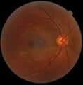
 Normal fundus photograph of the left eye, showing macula and optic disc
 -
 Digital retinography for retina and macula evaluation
 -
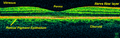 OCT scan of retina, displaying layers including macula
 -
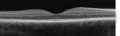 Spectral-domain OCT of the macula, cross-sectional view
 -
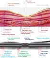 Histological and OCT image comparison of macular tissue


See Also[edit]
- Retina
- Fovea
- Photoreceptor cells
- Visual acuity
- Ophthalmology
- Macular degeneration
- Age-related macular degeneration
- Optical coherence tomography
| Anatomy of the globe of the human eye | ||||||||||||||||||
|---|---|---|---|---|---|---|---|---|---|---|---|---|---|---|---|---|---|---|
|


Medical Disclaimer: WikiMD is for informational purposes only and is not a substitute for professional medical advice. Content may be inaccurate or outdated and should not be used for diagnosis or treatment. Always consult your healthcare provider for medical decisions. Verify information with trusted sources such as CDC.gov and NIH.gov. By using this site, you agree that WikiMD is not liable for any outcomes related to its content. See full disclaimer.
Credits:Most images are courtesy of Wikimedia commons, and templates, categories Wikipedia, licensed under CC BY SA or similar.
Translate page: - East Asian
中文,
日本,
한국어,
South Asian
हिन्दी,
தமிழ்,
తెలుగు,
Urdu,
ಕನ್ನಡ,
Southeast Asian
Indonesian,
Vietnamese,
Thai,
မြန်မာဘာသာ,
বাংলা
European
español,
Deutsch,
français,
Greek,
português do Brasil,
polski,
română,
русский,
Nederlands,
norsk,
svenska,
suomi,
Italian
Middle Eastern & African
عربى,
Turkish,
Persian,
Hebrew,
Afrikaans,
isiZulu,
Kiswahili,
Other
Bulgarian,
Hungarian,
Czech,
Swedish,
മലയാളം,
मराठी,
ਪੰਜਾਬੀ,
ગુજરાતી,
Portuguese,
Ukrainian
