Orbit (anatomy)
The Orbit is the cavity or socket of the skull in which the eye and its appendages are situated. The orbit is formed by the cheekbone, the forehead, the temple, and the side of the nose. The eye is cushioned within the orbit by pads of fat.
Structure[edit]
The orbit has a volume of 30 millilitres, and a height, width, and depth of approximately 40, 35, and 45 millimetres respectively. The entrance to the orbital cavity is referred to as the base, and the back as the apex. Four bones contribute to the formation of the orbital cavity.
Bones[edit]
The bones of the orbit include:
- The frontal bone
- The zygomatic bone
- The ethmoid bone
- The sphenoid bone
- The maxillary bone
- The palatine bone
- The lacrimal bone
Function[edit]
The primary function of the orbit is to provide a cavity in which the eye and its appendages are housed. This protects the eye from injury and allows for the movement of the eye.
Clinical significance[edit]
Conditions that can affect the orbit and therefore the eye include Orbital cellulitis, Orbital fracture, and Graves' disease.
See also[edit]
References[edit]
| Anatomy of the globe of the human eye | ||||||||||||||||||
|---|---|---|---|---|---|---|---|---|---|---|---|---|---|---|---|---|---|---|
|
| Neurocranium of the skull | ||||||||||||||||||||||||||||||||
|---|---|---|---|---|---|---|---|---|---|---|---|---|---|---|---|---|---|---|---|---|---|---|---|---|---|---|---|---|---|---|---|---|
|

This WikiMD article can only be edited by registered and verified editors. You can log in or register.
-
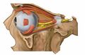
 Anterior view of the eye orbit anatomy
 -
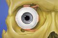
 3D model of the orbit of the face
 -
 Diagram of the orbital bones
 -
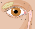 Diagram of the tear system
 -
 Orbit (anatomy)
 -
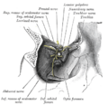
 Anatomy of the orbit from Gray's Anatomy
 -
 Muscles of the orbit from Gray's Anatomy
 -
 Orbit (anatomy)
 -
 Lateral view of the orbit showing nerves
 -
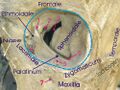
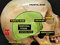 Temporal fossa related to the orbit


")
Medical Disclaimer: WikiMD is for informational purposes only and is not a substitute for professional medical advice. Content may be inaccurate or outdated and should not be used for diagnosis or treatment. Always consult your healthcare provider for medical decisions. Verify information with trusted sources such as CDC.gov and NIH.gov. By using this site, you agree that WikiMD is not liable for any outcomes related to its content. See full disclaimer.
Credits:Most images are courtesy of Wikimedia commons, and templates, categories Wikipedia, licensed under CC BY SA or similar.
Translate page: - East Asian
中文,
日本,
한국어,
South Asian
हिन्दी,
தமிழ்,
తెలుగు,
Urdu,
ಕನ್ನಡ,
Southeast Asian
Indonesian,
Vietnamese,
Thai,
မြန်မာဘာသာ,
বাংলা
European
español,
Deutsch,
français,
Greek,
português do Brasil,
polski,
română,
русский,
Nederlands,
norsk,
svenska,
suomi,
Italian
Middle Eastern & African
عربى,
Turkish,
Persian,
Hebrew,
Afrikaans,
isiZulu,
Kiswahili,
Other
Bulgarian,
Hungarian,
Czech,
Swedish,
മലയാളം,
मराठी,
ਪੰਜਾਬੀ,
ગુજરાતી,
Portuguese,
Ukrainian
