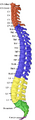
The Cervical Vertebræ
Anatomy > Gray's Anatomy of the Human Body > II. Osteology > The Cervical vertebrae.
Cervical Vertebrae[edit]
In humans, cervical vertebrae are the smallest of the true vertebrae and can be readily distinguished from those of the thoracic or lumbar regions by the presence of a foramen (hole) in each transverse process, through which the vertebral artery, vertebral veins, and inferior cervical ganglion pass. The remainder of this article focuses upon human anatomy.
Structure[edit]

By convention, the cervical vertebrae are numbered, with the first one (C1) closest to the skull and higher numbered vertebrae (C2–C7) proceeding away from the skull and down the spine. The general characteristics of the third through sixth cervical vertebrae are described here. The first, second, and seventh vertebrae are extraordinary, and are detailed later.
- The bodies of these four vertebrae are small, and broader from side to side than from front to back.
- The anterior and posterior surfaces are flattened and of equal depth; the former is placed on a lower level than the latter, and its inferior border is prolonged downward, so as to overlap the upper and forepart of the vertebra below.
- The upper surface is concave transversely, and presents a projecting lip on either side.
- The lower surface is concave from front to back, convex from side to side, and presents laterally shallow concavities that receive the corresponding projecting lips of the underlying vertebra.
- The pedicles are directed laterally and backward, and attach to the body midway between its upper and lower borders, so that the superior vertebral notch is as deep as the inferior, but it is, at the same time, narrower.
- The laminae are narrow and thinner above than below; the vertebral foramen is large and of a triangular form.
- The spinous process is short and bifid, the two divisions being often of unequal size. Because the spinous processes are so short, certain superficial muscles (the trapezius and splenius capitis) attach to the nuchal ligament rather than directly to the vertebrae; the nuchal ligament itself attaching to the spinous processes of C2–C7 and to the posterior tubercle of the atlas.
- The superior and inferior articular processes of cervical vertebrae have fused on either or both sides to form articular pillars, columns of bone that project laterally from the junction of the pedicle and lamina.
- The articular facets are flat and of an oval form:
- the superior face backward, upward, and slightly medially.
- the inferior face forward, downward, and slightly laterally.
- The transverse processes are each pierced by the foramen transversarium, which, in the upper six vertebrae, gives passage to the vertebral artery and vein, as well as a plexus of sympathetic nerves. Each process consists of an anterior and a posterior part. These two parts are joined, outside the foramen, by a bar of bone that exhibits a deep sulcus on its upper surface for the passage of the corresponding spinal nerve.
- The anterior portion is the homologue of the rib in the thoracic region, and is therefore named the costal process or costal element. It arises from the side of the body, is directed laterally in front of the foramen, and ends in a tubercle, the anterior tubercle.
- The posterior part, the true transverse process, springs from the vertebral arch behind the foramen and is directed forward and laterally; it ends in a flattened vertical tubercle, the posterior tubercle.
The anterior tubercle of the sixth cervical vertebra is known as the carotid tubercle or Chassaignac tubercle. This separates the carotid artery from the vertebral artery and the carotid artery can be massaged against this tubercle to relieve the symptoms of supraventricular tachycardia. The carotid tubercle is also used as a landmark for anaesthesia of the brachial plexus and cervical plexus.
The cervical spinal nerves emerge from above the cervical vertebrae. For example, the cervical spinal nerve 3 (C3) passes above C3.
Atlas and axis[edit]
The atlas (C1) and axis (C2) are the two topmost vertebrae.
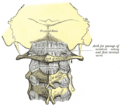
The atlas (C1) is the topmost vertebra, and along with the axis forms the joint connecting the skull and spine. It lacks a vertebral body, spinous process, and discs either superior or inferior to it. It is ring-like and consists of an anterior arch, posterior arch, and two lateral masses.
The axis (C2) forms the pivot on which the atlas rotates. The most distinctive characteristic of this bone is the strong odontoid process (dens) that rises perpendicularly from the upper surface of the body and articulates with C1. The body is deeper in front than behind, and prolonged downward anteriorly so as to overlap the upper and front part of the third vertebra.
Vertebra prominens[edit]

The vertebra prominens, or C7, has a distinctive long and prominent spinous process, which is palpable from the skin surface. Sometimes, the seventh cervical vertebra is associated with an abnormal extra rib, known as a cervical rib, which develops from the anterior root of the transverse process. These ribs are usually small, but may occasionally compress blood vessels (such as the subclavian artery or subclavian vein) or nerves in the brachial plexus, causing pain, numbness, tingling, and weakness in the upper limb, a condition known as thoracic outlet syndrome. Very rarely, this rib occurs in a pair.
The long spinous process of C7 is thick and nearly horizontal in direction. It is not bifurcated, and ends in a tubercle that the ligamentum nuchae attaches to. This process is not always the most prominent of the spinous processes, being found only about 70% of the time, C6 or T1 can sometimes be the most prominent.
The transverse processes are of considerable size; their posterior roots are large and prominent, while the anterior are small and faintly marked. The upper surface of each usually has a shallow sulcus for the eighth spinal nerve, and its extremity seldom presents more than a trace of bifurcation.
The transverse foramen may be as large as that in the other cervical vertebrae, but it is generally smaller on one or both sides; occasionally, it is double, and sometimes it is absent.
On the left side, it occasionally gives passage to the vertebral artery; more frequently, the vertebral vein traverses it on both sides, but the usual arrangement is for both artery and vein to pass in front of the transverse process, not through the foramen.
Function[edit]
The movement of nodding the head takes place predominantly through flexion and extension at the atlanto-occipital joint between the atlas and the occipital bone. However, the cervical spine is comparatively mobile, and some component of this movement is due to flexion and extension of the vertebral column itself. This movement between the atlas and occipital bone is often referred to as the "yes joint", owing to its nature of being able to move the head in an up-and-down fashion.
The movement of shaking or rotating the head left and right happens almost entirely at the joint between the atlas and the axis, the atlanto-axial joint. A small amount of rotation of the vertebral column itself contributes to the movement. This movement between the atlas and axis is often referred to as the "no joint", owing to its nature of being able to rotate the head in a side-to-side fashion.
Additional images[edit]
-
-
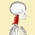 Position of cervical vertebrae (shown in red). Animation.
 -
Illustration of cervical vertebrae
-
 Shape of cervical vertebrae (shown in blue and yellow). Animation.
 -
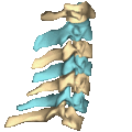
Cervical vertebrae, lateral view (shown in blue and yellow)
-
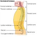 Vertebral column
 -
 Vertebral column
 -
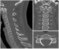
X-ray of cervical vertebrae
-
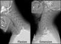 X-ray of cervical spine in flexion and extension
 -
 First cervical vertebra, or atlas
 -
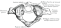
 Second cervical vertebra, or epistropheus, from above
 -
 Second cervical vertebra, epistropheus, or axis, from the side
 -
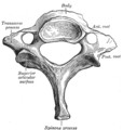 Seventh cervical vertebra
 -
 Posterior atlanto-occipital membrane and atlantoaxial ligament
 -
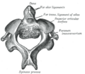
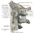 Median sagittal section through the occipital bone and first three cervical vertebrae
 -
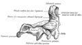
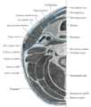 Section of the neck at about the level of the sixth cervical vertebra
 -
Anterior view of cervical spine showing the vertebral arteries along with the spinal nerves. See this in 3d here.
.jpg)


External links[edit]
- Diagram at kenyon.edu
- Cervical Spine Anatomy
- Mnemonic for Landmarks
- Cervical vertebra quiz
- Cervival vertebrae - BlueLink Anatomy - University of Michigan Medical School
| Bones of the torso | ||||||||||||||||
|---|---|---|---|---|---|---|---|---|---|---|---|---|---|---|---|---|
|
| Spinal nerves | ||||||||||
|---|---|---|---|---|---|---|---|---|---|---|
|
Gray's Anatomy[edit]
- Gray's Anatomy Contents
- Gray's Anatomy Subject Index
- About Classic Gray's Anatomy
- Glossary of anatomy terms
Anatomy atlases (external)[edit]
[1] - Anatomy Atlases
| Human systems and organs | ||||||||||||||
|---|---|---|---|---|---|---|---|---|---|---|---|---|---|---|
|
- ↑ David M., Principles of Life. online version, Palgrave Macmillan, ISBN 978-1-4641-6298-5, Pages: 280–,

Medical Disclaimer: WikiMD is for informational purposes only and is not a substitute for professional medical advice. Content may be inaccurate or outdated and should not be used for diagnosis or treatment. Always consult your healthcare provider for medical decisions. Verify information with trusted sources such as CDC.gov and NIH.gov. By using this site, you agree that WikiMD is not liable for any outcomes related to its content. See full disclaimer.
Credits:Most images are courtesy of Wikimedia commons, and templates, categories Wikipedia, licensed under CC BY SA or similar.
Translate page: - East Asian
中文,
日本,
한국어,
South Asian
हिन्दी,
தமிழ்,
తెలుగు,
Urdu,
ಕನ್ನಡ,
Southeast Asian
Indonesian,
Vietnamese,
Thai,
မြန်မာဘာသာ,
বাংলা
European
español,
Deutsch,
français,
Greek,
português do Brasil,
polski,
română,
русский,
Nederlands,
norsk,
svenska,
suomi,
Italian
Middle Eastern & African
عربى,
Turkish,
Persian,
Hebrew,
Afrikaans,
isiZulu,
Kiswahili,
Other
Bulgarian,
Hungarian,
Czech,
Swedish,
മലയാളം,
मराठी,
ਪੰਜਾਬੀ,
ગુજરાતી,
Portuguese,
Ukrainian
