The Lacrimal Bone
Anatomy > Gray's Anatomy of the Human Body > II. [Osteology]] > 5b. 3. The Lacrimal Bone
Henry Gray (1821–1865). Anatomy of the Human Body. 1918.
The Lacrimal Bone[edit]
(Os Lacrimale)
The lacrimal bone the smallest and most fragile bone of the face, is situated at the front part of the medial wall of the orbit (Fig. 164). It has two surfaces and four borders.
Surfaces[edit]
The lateral or orbital surface (Fig. 163) is divided by a vertical ridge, the posterior lacrimal crest into two parts. In front of this crest is a longitudinal groove, the lacrimal sulcus (sulcus lacrimalis), the inner margin of which unites with the frontal process of the maxilla, and the lacrimal fossa is thus completed. The upper part of this fossa lodges the lacrimal sac, the lower part, the naso-lacrimal duct.
The portion behind the crest is smooth, and forms part of the medial wall of the orbit. The crest, with a part of the orbital surface immediately behind it, gives origin to the lacrimal part of the Orbicularis oculi and ends below in a small, hook-like projection, the lacrimal hamulus which articulates with the lacrimal tubercle of the maxilla, and completes the upper orifice of the lacrimal canal; it sometimes exists as a separate piece, and is then called the lesser lacrimal bone
The medial or nasal surface presents a longitudinal furrow, corresponding to the crest on the lateral surface. The area in front of this furrow forms part of the middle meatus of the nose; that behind it articulates with the ethmoid, and completes some of the anterior ethmoidal cells.

Borders[edit]
Of the four borders the anterior articulates with the frontal process of the maxilla; the posterior with the lamina papyracea of the ethmoid; the superior with the frontal bone.
The inferior is divided by the lower edge of the posterior lacrimal crest into two parts: the posterior part articulates with the orbital plate of the maxilla; the anterior is prolonged downward as the descending process which articulates with the lacrimal process of the inferior nasal concha, and assists in forming the canal for the nasolacrimal duct.
Ossification[edit]
The lacrimal is ossified from a single center, which appears about the twelfth week in the membrane covering the cartilaginous nasal capsule.
Articulations[edit]
The lacrimal articulates with four bones: two of the cranium, the frontal and ethmoid, and two of the face, the maxilla and the inferior nasal concha.
Additional images[edit]
-
 Position of the lacrimal bones (shown in green). Animation.
 -
 Animation. Some bones are removed to show the position of the lacrimal bones (shown in green).
 -
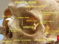
 Orbital bones. Lacrimal bone shown in green.
 -
 A left lacrimal bone. Enlarged. Animation.
 -
 Lacrimal bone
 -
 Lacrimal bone


External links[edit]
- Atlas image: eye_5 at the University of Michigan Health System
- Atlas image: rsa2p4 at the University of Michigan Health System
| The orbit of the eye | ||||||||||
|---|---|---|---|---|---|---|---|---|---|---|
|
| The facial skeleton of the skull | ||||||||||||||||||||||||
|---|---|---|---|---|---|---|---|---|---|---|---|---|---|---|---|---|---|---|---|---|---|---|---|---|
|
Gray's Anatomy[edit]
- Gray's Anatomy Contents
- Gray's Anatomy Subject Index
- About Classic Gray's Anatomy
- Glossary of anatomy terms
Anatomy atlases (external)[edit]
[1] - Anatomy Atlases
| Human systems and organs | ||||||||||||||
|---|---|---|---|---|---|---|---|---|---|---|---|---|---|---|
|

Medical Disclaimer: WikiMD is for informational purposes only and is not a substitute for professional medical advice. Content may be inaccurate or outdated and should not be used for diagnosis or treatment. Always consult your healthcare provider for medical decisions. Verify information with trusted sources such as CDC.gov and NIH.gov. By using this site, you agree that WikiMD is not liable for any outcomes related to its content. See full disclaimer.
Credits:Most images are courtesy of Wikimedia commons, and templates, categories Wikipedia, licensed under CC BY SA or similar.
Translate page: - East Asian
中文,
日本,
한국어,
South Asian
हिन्दी,
தமிழ்,
తెలుగు,
Urdu,
ಕನ್ನಡ,
Southeast Asian
Indonesian,
Vietnamese,
Thai,
မြန်မာဘာသာ,
বাংলা
European
español,
Deutsch,
français,
Greek,
português do Brasil,
polski,
română,
русский,
Nederlands,
norsk,
svenska,
suomi,
Italian
Middle Eastern & African
عربى,
Turkish,
Persian,
Hebrew,
Afrikaans,
isiZulu,
Kiswahili,
Other
Bulgarian,
Hungarian,
Czech,
Swedish,
മലയാളം,
मराठी,
ਪੰਜਾਬੀ,
ગુજરાતી,
Portuguese,
Ukrainian
