The Maxillæ (Upper Jaw)
Anatomy > Gray's Anatomy of the Human Body > II. [Osteology]] > 5b. The Facial Bones. 2. The Maxillae
Henry Gray (1821–1865). Anatomy of the Human Body. 1918.
The Maxilla[edit]
The maxilla (plural: maxillae )[1] in vertebrates is the upper fixed (not fixed in Neopterygii) bone of the jaw formed from the fusion of two maxillary bones. The upper jaw includes the hard palate in the front of the mouth.[2][3] The two maxillary bones are fused at the intermaxillary suture, forming the anterior nasal spine. This is similar to the mandible (lower jaw), which is also a fusion of two mandibular bones at the mandibular symphysis. The mandible is the movable part of the jaw.
Structure[edit]

In humans, the maxilla consists of:
- The body of the maxilla
- Four processes
- The zygomatic process
- The frontal process of maxilla
- The alveolar process
- The palatine process
- Three surfaces - anterior, posterior, medial
- The Infraorbital foramen
- The maxillary sinus
- The incisive foramen
Articulations[edit]
Each maxilla articulates with nine bones:
- two of the cranium: the frontal and ethmoid
- seven of the face: the nasal, zygomatic, lacrimal, inferior nasal concha, palatine, vomer, and the adjacent fused maxilla.
Sometimes it articulates with the orbital surface, and sometimes with the lateral pterygoid plate of the sphenoid.
Development[edit]


The maxilla is ossified in membrane. Mall and Fawcett maintain that it is ossified from two centers only, one for the maxilla proper and one for the premaxilla.[4][5]
These centers appear during the sixth week of prenatal development and unite in the beginning of the third month, but the suture between the two portions persists on the palate until nearly middle life. Mall states that the frontal process is developed from both centers.
The maxillary sinus appears as a shallow groove on the nasal surface of the bone about the fourth month of development, but does not reach its full size until after the second dentition.
The maxilla was formerly described as ossifying from six centers, viz.,
- one, the orbitonasal, forms that portion of the body of the bone which lies medial to the infraorbital canal, including the medial part of the floor of the orbit and the lateral wall of the nasal cavity;
- a second, the zygomatic, gives origin to the portion which lies lateral to the infraorbital canal, including the zygomatic process;
- from a third, the palatine, is developed the palatine process posterior to the incisive canal together with the adjoining part of the nasal wall;
- a fourth, the premaxillary, forms the incisive bone which carries the incisor teeth and corresponds to the premaxilla of the lower vertebrates;
- a fifth, the nasal, gives rise to the frontal process and the portion above the canine tooth;
- and a sixth, the infravomerine, lies between the palatine and premaxillary centers and beneath the vomer; this center, together with the corresponding center of the opposite bone, separates the incisive canals from each other.
Function[edit]

The alveolar process of the maxillae holds the upper teeth, and is referred to as the maxillary arch. Each maxilla attaches laterally to the zygomatic bones (cheek bones).
Each maxilla assists in forming the boundaries of three cavities:
- the roof of the mouth
- the floor and lateral wall of the nasal cavity
- the wall of the orbit
Each maxilla also enters into the formation of two fossae: the infratemporal and pterygopalatine, and two fissures, the inferior orbital and pterygomaxillary. -When the tender bones of the upper jaw and lower nostril are severely or repetitively damaged, at any age the surrounding cartilage can begin to deteriorate just as it does after death. (January 2016)
Additional images[edit]
-
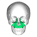 Skull. Maxilla shown in green.

External links[edit]
- Anatomy photo:22:os-1901 at the SUNY Downstate Medical Center
| The orbit of the eye | ||||||||||
|---|---|---|---|---|---|---|---|---|---|---|
|
| The facial skeleton of the skull | ||||||||||||||||||||||||
|---|---|---|---|---|---|---|---|---|---|---|---|---|---|---|---|---|---|---|---|---|---|---|---|---|
|
Gray's Anatomy[edit]
- Gray's Anatomy Contents
- Gray's Anatomy Subject Index
- About Classic Gray's Anatomy
- Glossary of anatomy terms
Anatomy atlases (external)[edit]
[1] - Anatomy Atlases
| Human systems and organs | ||||||||||||||
|---|---|---|---|---|---|---|---|---|---|---|---|---|---|---|
|
- ↑ OED 2nd edition, 1989.
- ↑ Merriam-Webster Online Dictionary.
- ↑ , Illustrated Anatomy of the Head and Neck, Elsevier, 2012, ISBN 978-1-4377-2419-6,
- ↑ Mall, Franklin P.."On ossification centers in human embryos less than one hundred days old".American Journal of Anatomy.1906;5(4)
- 433–458.doi:10.1002/aja.1000050403.Full text.
- ↑ Fawcett, Edward."Some Notes on the Epiphyses of the Ribs".Journal of Anatomy and Physiology.1911;45(Pt 2)
")
Medical Disclaimer: WikiMD is for informational purposes only and is not a substitute for professional medical advice. Content may be inaccurate or outdated and should not be used for diagnosis or treatment. Always consult your healthcare provider for medical decisions. Verify information with trusted sources such as CDC.gov and NIH.gov. By using this site, you agree that WikiMD is not liable for any outcomes related to its content. See full disclaimer.
Credits:Most images are courtesy of Wikimedia commons, and templates, categories Wikipedia, licensed under CC BY SA or similar.
Translate page: - East Asian
中文,
日本,
한국어,
South Asian
हिन्दी,
தமிழ்,
తెలుగు,
Urdu,
ಕನ್ನಡ,
Southeast Asian
Indonesian,
Vietnamese,
Thai,
မြန်မာဘာသာ,
বাংলা
European
español,
Deutsch,
français,
Greek,
português do Brasil,
polski,
română,
русский,
Nederlands,
norsk,
svenska,
suomi,
Italian
Middle Eastern & African
عربى,
Turkish,
Persian,
Hebrew,
Afrikaans,
isiZulu,
Kiswahili,
Other
Bulgarian,
Hungarian,
Czech,
Swedish,
മലയാളം,
मराठी,
ਪੰਜਾਬੀ,
ગુજરાતી,
Portuguese,
Ukrainian
