The Thoractic Duct
Anatomy > Gray's Anatomy of the Human Body > VIII. The Lymphatic System > 2. The Thoracic Duct
Henry Gray (1821–1865). Anatomy of the Human Body. 1918.
The Thoracic Duct[edit]
The thoracic duct (ductus thoracicus) (

) conveys the greater part of the lymph and chyle into the blood. It is the common trunk of all the lymphatic vessels of the body, excepting those on the right side of the head, neck, and thorax, and right upper extremity, the right lung, right side of the heart, and the convex surface of the liver.
In the adult it varies in length from 38 to 45 cm. and extends from the second lumbar vertebra to the root of the neck. It begins in the abdomen by a triangular dilatation, the cisterna chyli which is situated on the front of the body of the second lumbar vertebra, to the right side of and behind the aorta, by the side of the right crus of the diaphragm.
It enters the thorax through the aortic hiatus of the diaphragm, and ascends through the posterior mediastinal cavity between the aorta and azygos vein. Behind it in this region are the vertebral column, the right intercostal arteries, and the hemiazygos veins as they cross to open into the azygos vein; in front of it are the diaphragm, esophagus, and pericardium, the last being separated from it by a recess of the right pleural cavity.
Opposite the fifth thoracic vertebra, it inclines toward the left side, enters the superior mediastinal cavity, and ascends behind the aortic arch and the thoracic part of the left subclavian artery and between the left side of the esophagus and the left pleura, to the upper orifice of the thorax.
Passing into the neck it forms an arch which rises about 3 or 4 cm. above the clavicle and crosses anterior to the subclavian artery, the vertebral artery and vein, and the thyrocervical trunk or its branches.
It also passes in front of the phrenic nerve and the medial border of the Scalenus anterior, but is separated from these two structures by the prevertebral fascia. In front of it are the left common carotid artery, vagus nerve, and internal jugular vein; it ends by opening into the angle of junction of the left subclavian vein with the left internal jugular vein.
The thoracic duct, at its commencement, is about equal in diameter to a goose-quill, but it diminishes considerably in caliber in the middle of the thorax, and is again dilated just before its termination.
It is generally flexuous, and constricted at intervals so as to present a varicose appearance. Not infrequently it divides in the middle of its course into two vessels of unequal size which soon reunite, or into several branches which form a plexiform interlacement.
It occasionally divides at its upper part into two branches, right and left; the left ending in the usual manner, while the right opens into the right subclavian vein, in connection with the right lymphatic duct. The thoracic duct has several valves; at its termination it is provided with a pair, the free borders of which are turned toward the vein, so as to prevent the passage of venous blood into the duct.
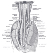
The cisterna chyli[edit]
The cisterna chyli (receptaculum chyli) (

) receives the two lumbar lymphatic trunks, right and left, and the intestinal lymphatic trunk. The lumbar trunks are formed by the union of the efferent vessels from the lateral aortic lymph glands. They receive the lymph from the lower limbs, from the walls and viscera of the pelvis, from the kidneys and suprarenal gland and the deep lymphatics of the greater part of the abdominal wall. The intestinal trunk receives the lymph from the stomach and intestine, from the pancreas and spleen, and from the lower and front part of the liver.
Tributaries[edit]
Opening into the commencement of the thoracic duct, on either side, is a descending trunk from the posterior intercostal lymph glands of the lower six or seven intercostal spaces.
In the thorax the duct is joined, on either side, by a trunk which drains the upper lumbar lymph gland and pierces the crus of the diaphragm. It also receives the efferents from the posterior mediastinal lymph glands and from the posterior intercostal lymph glands of the upper six left spaces.
In the neck it is joined by the left jugular and left subclavian trunks and sometimes by the left bronchomediastinal trunk the last-named, however, usually opens independently into the junction of the left subclavian and internal jugular veins.

FIG. 601– Terminal collecting trunks of right side. 'a' Jugular trunk. 'b' Subclavian trunk. c Bronchomediastinal trunk. d Right lymphatic trunk. e Gland of internal mammary chain. f Gland of deep cervical chain. (Poirier and Charpy.) (Picture From the Classic Gray's Anatomy)
Function[edit]
The thoracic duct collects most of the lymph in the body other than from the right thorax, arm, head, and neck which are drained by the right lymphatic duct.

The lymph transport, in the thoracic duct, is mainly caused by the action of breathing, aided by the duct's smooth muscle and by internal valves which prevent the lymph from flowing back down again. There are also two valves at the junction of the duct with the left subclavian vein, to prevent the flow of venous blood into the duct. In adults, the thoracic duct transports up to 4 L of lymph per day.
Additional images[edit]
-
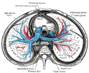 Transverse section of thorax, showing relations of pulmonary artery.
 -
 The arch of the aorta, and its branches.
 -
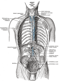 Deep lymph nodes and vessels of the thorax and abdomen (diagrammatic).
 -
 The position and relation of the esophagus in the cervical region and in the posterior mediastinum. Seen from behind.
 -
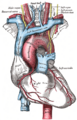
 Front photo of the Ductus Thoracicus in the Human mediastinum with the heart and part of the pericard removed.


External links[edit]
- Anatomy figure: 21:05-02 at Human Anatomy Online, SUNY Downstate Medical Center — "The thoracic duct and azygos venous network"
- Anatomy image:8901 at the SUNY Downstate Medical Center
- figures/chapter_24/24-5.HTM: Basic Human Anatomy at Dartmouth Medical School
| Anatomy of the lymphatic system | ||||||||||||||||||||||||||||||||
|---|---|---|---|---|---|---|---|---|---|---|---|---|---|---|---|---|---|---|---|---|---|---|---|---|---|---|---|---|---|---|---|---|
|
Gray's Anatomy[edit]
- Gray's Anatomy Contents
- Gray's Anatomy Subject Index
- About Classic Gray's Anatomy
- Glossary of anatomy terms
Anatomy atlases (external)[edit]
[1] - Anatomy Atlases
| Human systems and organs | ||||||||||||||
|---|---|---|---|---|---|---|---|---|---|---|---|---|---|---|
|

Medical Disclaimer: WikiMD is for informational purposes only and is not a substitute for professional medical advice. Content may be inaccurate or outdated and should not be used for diagnosis or treatment. Always consult your healthcare provider for medical decisions. Verify information with trusted sources such as CDC.gov and NIH.gov. By using this site, you agree that WikiMD is not liable for any outcomes related to its content. See full disclaimer.
Credits:Most images are courtesy of Wikimedia commons, and templates, categories Wikipedia, licensed under CC BY SA or similar.
Translate page: - East Asian
中文,
日本,
한국어,
South Asian
हिन्दी,
தமிழ்,
తెలుగు,
Urdu,
ಕನ್ನಡ,
Southeast Asian
Indonesian,
Vietnamese,
Thai,
မြန်မာဘာသာ,
বাংলা
European
español,
Deutsch,
français,
Greek,
português do Brasil,
polski,
română,
русский,
Nederlands,
norsk,
svenska,
suomi,
Italian
Middle Eastern & African
عربى,
Turkish,
Persian,
Hebrew,
Afrikaans,
isiZulu,
Kiswahili,
Other
Bulgarian,
Hungarian,
Czech,
Swedish,
മലയാളം,
मराठी,
ਪੰਜਾਬੀ,
ગુજરાતી,
Portuguese,
Ukrainian
