Arches of the Foot
Anatomical structure and function of the foot arches
Arches of the Foot are structural features formed by the tarsal bones, metatarsal bones, and supported by ligaments, tendons, and intrinsic muscles of the foot. They provide strength and flexibility, allowing the foot to support the weight of the human body during standing, walking, and running.
The bony architecture of the foot is arranged into multiple arches to maximize support with minimal material. These arches distribute body weight and provide shock absorption. There are two main types:
- Longitudinal arches – running lengthwise
- Transverse arches – running across the foot
Longitudinal Arches[edit]
The longitudinal arches are the most prominent and are divided into:
Medial Longitudinal Arch[edit]
The medial longitudinal arch is the higher and more elastic of the two. It is composed of the following bones:
- Calcaneus
- Talus
- Navicular bone
- Three cuneiform bones
- First, second, and third metatarsals
The highest point is the superior articular surface of the talus. The posterior end rests on the tuberosity of the calcaneus, while the anterior end is supported by the heads of the first three metatarsals.
The stability and elasticity of this arch are maintained by:
- Plantar calcaneonavicular ligament (spring ligament)
- Deltoid ligament
- Tibialis posterior muscle
- Tibialis anterior muscle
- Peroneus longus muscle
- Plantar aponeurosis
- Intrinsic muscles of the foot
The joint most susceptible to collapse is the talonavicular joint, which is reinforced by the spring ligament and the fan-like insertion of the tibialis posterior tendon.
Lateral Longitudinal Arch[edit]
The lateral longitudinal arch is flatter and more rigid. It comprises:
- Calcaneus
- Cuboid bone
- Fourth and fifth metatarsals
The apex lies at the calcaneocuboid joint, known for its locking mechanism, allowing limited motion. This arch is stabilized by:
- Long plantar ligament
- Plantar calcaneocuboid ligament (short plantar)
- Extensor digitorum longus muscle
- Flexor digitorum brevis muscle
- Abductor digiti minimi muscle
While the medial and lateral arches are often discussed separately, functionally they contribute to the unified longitudinal arch of the foot. Notably, a core segment of the longitudinal arch includes the cuboid, third cuneiform, and third metatarsal.
Transverse Arches[edit]
The foot also exhibits a series of transverse arches, particularly at:
- The base of the metatarsal bones
- The cuboid and cuneiform bones region
These arches form semi-domes with their concavities directed inferiorly and medially. When both feet are placed together, they form a complete dome.
Stabilization of the transverse arches involves:
- Interosseous ligaments
- Plantar ligaments
- Dorsal ligaments
- Adductor hallucis muscle (transverse head)
- Peroneus longus muscle (crosses the foot and supports the arch)
Function[edit]
The arches of the foot are essential in:
- Supporting body weight in various postures
- Absorbing impact during locomotion
- Distributing pressure evenly
- Assisting in propulsion during gait
The medial arch specifically accommodates soft tissue structures such as the plantar aponeurosis, which acts as a spring to absorb and release energy, making gait more efficient and protecting the musculoskeletal system from overuse injuries.
Clinical Significance[edit]
- Flattening of the arches results in pes planus (flat feet), which may cause pain or impaired gait.
- Exaggeration results in pes cavus, leading to instability or increased risk of injury.
- The arches are tested during physical examination and gait analysis and may be supported with orthotic devices.
Additional Images[edit]
-
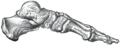 Skeleton of foot - medial aspect
 -
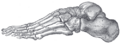 Skeleton of foot - lateral aspect
Bibliography[edit]
- R. Fick: Handbuch der Anatomie und Mechanik der Gelenke (Bardeleben’s Handbuch der Anatomie)
External Links[edit]
- Overview at gla.ac.uk
- Arches of the Foot – Archive
- Arches diagram
| Joints and ligaments of the human leg | ||||||||||||||
|---|---|---|---|---|---|---|---|---|---|---|---|---|---|---|
|
Gray's Anatomy[edit]
- Gray's Anatomy Contents
- Gray's Anatomy Subject Index
- About Classic Gray's Anatomy
- Glossary of anatomy terms
Anatomy atlases (external)[edit]
[1] - Anatomy Atlases
| Human systems and organs | ||||||||||||||
|---|---|---|---|---|---|---|---|---|---|---|---|---|---|---|
|
Adapted from the Classic Grays Anatomy of the Human Body 1918 edition (public domain)

Medical Disclaimer: WikiMD is for informational purposes only and is not a substitute for professional medical advice. Content may be inaccurate or outdated and should not be used for diagnosis or treatment. Always consult your healthcare provider for medical decisions. Verify information with trusted sources such as CDC.gov and NIH.gov. By using this site, you agree that WikiMD is not liable for any outcomes related to its content. See full disclaimer.
Credits:Most images are courtesy of Wikimedia commons, and templates, categories Wikipedia, licensed under CC BY SA or similar.
Translate page: - East Asian
中文,
日本,
한국어,
South Asian
हिन्दी,
தமிழ்,
తెలుగు,
Urdu,
ಕನ್ನಡ,
Southeast Asian
Indonesian,
Vietnamese,
Thai,
မြန်မာဘာသာ,
বাংলা
European
español,
Deutsch,
français,
Greek,
português do Brasil,
polski,
română,
русский,
Nederlands,
norsk,
svenska,
suomi,
Italian
Middle Eastern & African
عربى,
Turkish,
Persian,
Hebrew,
Afrikaans,
isiZulu,
Kiswahili,
Other
Bulgarian,
Hungarian,
Czech,
Swedish,
മലയാളം,
मराठी,
ਪੰਜਾਬੀ,
ગુજરાતી,
Portuguese,
Ukrainian
