Surface Markings of the Lower Extremity
Anatomy > Gray's Anatomy of the Human Body > XII. Surface Anatomy and Surface Markings > 14. Surface Markings of the Lower Extremity
Henry Gray (1821–1865). Anatomy of the Human Body. 1918.
Surface Markings of the Lower Extremity[edit | edit source]
Bony Landmarks[edit | edit source]
The anterior superior iliac spine is at the level of the sacral promontory—the posterior at the level of the spinous process of the second sacral vertebra. A horizontal line through the highest points of the iliac crests passes also through the spinous process of the fourth lumbar vertebra, while, as already pointed out (page 1315), the transtubercular plane through the tubercles on the iliac crests cuts the body of the fifth lumbar vertebra.
The upper margin of the greater sciatic notch is opposite the spinous process of the third sacral vertebra, and slightly below this level is the posterior inferior iliac spine. The surface markings of the posterior inferior iliac spine and the ischial spine are both situated in a line which joins the posterior superior iliac spine to the outer part of the ischial tuberosity; the posterior inferior spine is 5 cm. and the ischial spine 10 cm. below the posterior superior spine; the ischial spine is opposite the first piece of the coccyx. With the body in the erect posture the line joining the public tubercle to the top of the greater trochanter is practically horizontal; the middle of this line overlies the acetabulum and the head of the femur.
A line used for clinical purposes is that of Nélaton (Fig. 1243), which is drawn from the anterior superior iliac spine to the most prominent part of the ischial tuberosity; it crosses the center of the acetabulum and the upper border of the greater trochanter. Another surface marking of clinical importance is Bryant’s triangle which is mapped out thus: a line from the anterior superior iliac spine to the top of the greater trochanter forms the base of the triangle; its sides are formed respectively by a horizontal line from the anterior superior iliac spine and a vertical line from the top of the greater trochanter.
Articulations[edit | edit source]
The posterior superior iliac spine overlies the center of the sacroiliac articulations
The hip-joint may be indicated, as described above, by the center of a horizontal line from the pubic tubercle to the top of the greater trochanter; or more generally, it is below and slightly lateral to the middle of the inguinal ligament.
The knee-joint is superficial and requires no surface marking.
The level of the ankle-joint is that of a transverse line about 1 cm. above the level of the tip of the medial malleolus. If the foot be forcibly extended, the head of the talus appears as a rounded prominence on the medial side of the dorsum; just in front of this prominence and behind the tuberosity of the navicular is the talonavicular joint The calcaneocuboid joint is situated midway between the lateral malleolus and the prominent base of the fifth metatarsal bone; the line indicating it is parallel to that of the talonavicular joint.
The line of the fifth tarsometatarsal joint is very oblique; it starts from the projection of the base of the fifth metatarsal bone, and if continued would pass through the head of the first metatarsal. The lines of the fourth and third tarsometatarsal joints are less oblique. The first tarsometatarsal joint corresponds to a groove which can be felt by making firm pressure on the medial border of the foot 2.5 cm. in front of the tuberosity of the navicular bone; the position of the second tarsometatarsal joint is 1.25 cm. behind this.
The metatarsophalangeal joints are about 2.5 cm. behind the webs of the corresponding toes.
Muscles[edit | edit source]
None of the muscles require any special surface lines to indicate them, but there are three intermuscular spaces which occasionally require definition, viz., the femoral triangle, the adductor canal, and the popliteal fossa.
The femoral triangle is bounded above by the inguinal ligament, laterally by the medial border of Sartorius, and medially by the medial border of Adductor longus. In the triangle is the fossa ovalis, through which the great saphenous vein dips to join the femoral; the center of this fossa is about 4 cm. below and lateral to the pubic tubercle, its vertical diameter measures about 4 cm. and its transverse about 1.5 cm. The femoral ring is about 1.25 cm. lateral to the pubic tubercle.
The adductor canal occupies the medial part of the middle third of the thigh; it begins at the apex of the femoral triangle and lies deep to the vertical part of Sartorius. The popliteal fossa is bounded: above and medially by the tendons of Semimembranosus and Semitendinosus; above and laterally by the tendon of Biceps femoris; below and medially by the medial head of Gastrocnemius; below and laterally by the lateral head of Gastrocnemius and the Plantaris.
Mucous Sheaths[edit | edit source]
The positions of the mucous sheaths around the tendons about the ankle-joints are sufficiently indicated in Figs. 1241, 1242 (see also page 489).
Arteries[edit | edit source]
The points of emergence of the three main arteries on the buttock, viz., the superior and inferior gluteals and the internal pudendal, may be indicated in the following manner (Fig. 1244). With the femur slightly flexed and rotated inward, a line is drawn from the posterior superior iliac spine to the posterior superior angle of the greater trochanter; the point of emergence of the superior gluteal artery from the upper part of the greater sciatic foramen corresponds to the junction of the upper and middle thirds of this line.
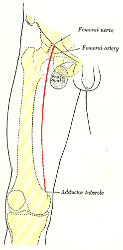
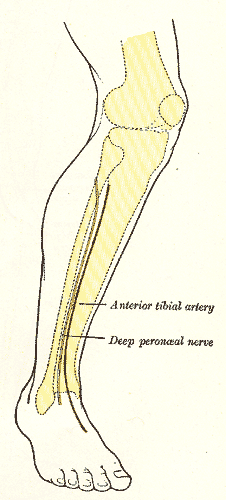
A second line is drawn from the posterior superior iliac spine to the outer part of the ischial tuberosity; the junction of its lower with its middle third marks the point of emergence of the inferior gluteal and internal pudendal arteries from the lower part of the greater sciatic foramen. The course of the femoral artery (Fig. 1245) is represented by the upper two-thirds of a line from a point midway between the anterior superior iliac spine and the symphysis pubis to the adductor tubercle, with the thigh abducted and rotated outward; the profunda femoris arises from it about 1 to 5 cm. below the inguinal ligament. The course of the upper part of the popliteal artery (Fig. 1247) is indicated by a line from the lateral margin of Semimembranosus at the junction of the middle and lower thirds of the thigh, obliquely downward to the middle of the popliteal fossa; from this point it runs vertically downward for about 2.5 cm. or to the level of a line through the lower part of the tibial tuberosity. The line indicating the anterior tibial artery (Fig. 1246) is drawn from the medial side of the head of the fibula to a point midway between the malleoli; the artery begins about 3 cm. below the head of the fibula.
The dorsalis pedis artery is represented on the dorsum of the foot by a line from the center of the interval between the malleoli to the proximal end of the first intermetatarsal space. The course of the posterior tibial artery (Fig. 1247) can be shown by a line from the end of the popliteal artery, i. e 2.5 cm. below the center of the popliteal fossa, to midway between the tip of the medial malleolus and the center of the convexity of the heel; its main branch, the peroneal artery begins about 7 or 8 cm. below the level of the knee-joint and follows the line of the fibula to the back of the lateral malleolus. The medial and lateral plantar arteries begin from the end of the posterior tibial; the medial extends to the middle of the plantar surface of the ball of the great toe, the lateral to within a finger’s breadth of the tuberosity of the fifth metatarsal bone; from this latter point the plantar arch crosses the foot transversely to the proximal end of the first intermetatarsal space.
Veins[edit | edit source]
The line of the great saphenous vein is from the front of the medial malleolus to the center of the fossa ovalis; the small saphenous vein runs from the back of the lateral malleolus to the center of the popliteal fossa.
Nerves[edit | edit source]
The course of the sciatic nerve (Fig. 1247) can be indicated by a line from a point midway between the outer border of the ischial tuberosity and the posterior superior angle of the greater trochanter to the upper angle of the popliteal fossa. The continuation of this line vertically through the center of the popliteal fossa represents the position of the tibial nerve while the common peroneal nerve follows the line of the tendon of Biceps femoris. The lines for the deep peroneal nerve and the continuation of the tibial nerve correspond respectively to those for the anterior and posterior tibial arteries.
Further reading[edit | edit source]
- ,
 The anatomical basis of medical practice,
 illustrated edition,
 Williams and Wilkins,
 1971,
- John S. P.,
 Surface Anatomy: The Anatomical Basis of Clinical Examination. online version,
 4th edition,
 Elsevier Health Sciences,


 ISBN 978-0-7020-4776-3,
- Johannes Wilhelm,
 Color Atlas of Anatomy – A Photographic Study of the Human Body. online version,
 7th edition,
 Wolters Kluwer Health/Lippincott Williams & Wilkins,
 2011,


 ISBN 978-1-58255-856-1,


 Pages: 204–5, 476–7,
- Joseph,
 Drawing the Living Figure,

 Dover Publications, Incorporated,
 1991,


 ISBN 0-486-26723-7,
| Anatomy and morphology | ||||||||||
|---|---|---|---|---|---|---|---|---|---|---|
|
| Human regional anatomy | ||||||||||
|---|---|---|---|---|---|---|---|---|---|---|
|
| Human surface anatomy and general regions | ||||||||||||
|---|---|---|---|---|---|---|---|---|---|---|---|---|
|
| Human surface anatomy and general regions | ||||||||||||
|---|---|---|---|---|---|---|---|---|---|---|---|---|
|
Gray's Anatomy[edit source]
- Gray's Anatomy Contents
- Gray's Anatomy Subject Index
- About Classic Gray's Anatomy
- Glossary of anatomy terms
Anatomy atlases (external)[edit source]
[1] - Anatomy Atlases
| This article is a medical stub. You can help WikiMD by expanding it! | |
|---|---|
| Human systems and organs | ||||||||||||||
|---|---|---|---|---|---|---|---|---|---|---|---|---|---|---|
|
Search WikiMD
Ad.Tired of being Overweight? Try W8MD's physician weight loss program.
Semaglutide (Ozempic / Wegovy and Tirzepatide (Mounjaro / Zepbound) available.
Advertise on WikiMD
|
WikiMD's Wellness Encyclopedia |
| Let Food Be Thy Medicine Medicine Thy Food - Hippocrates |
Translate this page: - East Asian
中文,
日本,
한국어,
South Asian
हिन्दी,
தமிழ்,
తెలుగు,
Urdu,
ಕನ್ನಡ,
Southeast Asian
Indonesian,
Vietnamese,
Thai,
မြန်မာဘာသာ,
বাংলা
European
español,
Deutsch,
français,
Greek,
português do Brasil,
polski,
română,
русский,
Nederlands,
norsk,
svenska,
suomi,
Italian
Middle Eastern & African
عربى,
Turkish,
Persian,
Hebrew,
Afrikaans,
isiZulu,
Kiswahili,
Other
Bulgarian,
Hungarian,
Czech,
Swedish,
മലയാളം,
मराठी,
ਪੰਜਾਬੀ,
ગુજરાતી,
Portuguese,
Ukrainian
Medical Disclaimer: WikiMD is not a substitute for professional medical advice. The information on WikiMD is provided as an information resource only, may be incorrect, outdated or misleading, and is not to be used or relied on for any diagnostic or treatment purposes. Please consult your health care provider before making any healthcare decisions or for guidance about a specific medical condition. WikiMD expressly disclaims responsibility, and shall have no liability, for any damages, loss, injury, or liability whatsoever suffered as a result of your reliance on the information contained in this site. By visiting this site you agree to the foregoing terms and conditions, which may from time to time be changed or supplemented by WikiMD. If you do not agree to the foregoing terms and conditions, you should not enter or use this site. See full disclaimer.
Credits:Most images are courtesy of Wikimedia commons, and templates, categories Wikipedia, licensed under CC BY SA or similar.
Contributors: Anish, Deepika vegiraju, Prab R. Tumpati, MD


