The Pancreas
Anatomy > Gray's Anatomy of the Human Body > XI. Splanchnology > 2j. The Pancreas
Henry Gray (1821–1865). Anatomy of the Human Body. 1918.
The Pancreas[edit]
The pancreas (Figs. 1097, 1098) is a compound racemose gland, analogous in its structures to the salivary glands, though softer and less compactly arranged than those organs. Its secretion, the pancreatic juice carried by the pancreatic duct to the duodenum, is an important digestive fluid. In addition the pancreas has an important internal secretion, probably elaborated by the cells of Langerhans, which is taken up by the blood stream and is concerned with sugar metabolism. It is long and irregularly prismatic in shape; its right extremity, being broad, is called the head and is connected to the main portion of the organ, or body by a slight constriction, the neck while its left extremity gradually tapers to form the tail It is situated transversely across the posterior wall of the abdomen, at the back of the epigastric and left hypochondriac regions. Its length varies from 12.5 to 15 cm., and its weight from 60 to 100 gm.


Relations[edit]
The Head[edit]
The Head (caput pancreatis) is flattened from before backward, and is lodged within the curve of the duodenum. Its upper border is overlapped by the superior part of the duodenum and its lower overlaps the horizontal part; its right and left borders overlap in front, and insinuate themselves behind, the descending and ascending parts of the duodenum respectively. The angle of junction of the lower and left lateral borders forms a prolongation, termed the uncinate process In the groove between the duodenum and the right lateral and lower borders in front are the anastomosing superior and inferior pancreaticoduodenal arteries; the common bile duct descends behind, close to the right border, to its termination in the descending part of the duodenum.
Anterior Surface The greater part of the right half of this surface is in contact with the transverse colon, only areolar tissue intervening. From its upper part the neck springs, its right limit being marked by a groove for the gastroduodenal artery. The lower part of the right half, below the transverse colon, is covered by peritoneum continuous with the inferior layer of the transverse mesocolon, and is in contact with the coils of the small intestine. The superior mesenteric artery passes down in front of the left half across the uncinate process; the superior mesenteric vein runs upward on the right side of the artery and, behind the neck, joins with the lienal vein to form the portal vein.
Posterior Surface The posterior surface is in relation with the inferior vena cava, the common bile duct, the renal veins, the right crus of the diaphragm, and the aorta.
The Neck[edit]
The Neck springs from the right upper portion of the front of the head. It is about 2.5 cm. long, and is directed at first upward and forward, and then upward and to the left to join the body; it is somewhat flattened from above downward and backward. Its antero-superior surface supports the pylorus; its postero-inferior surface is in relation with the commencement of the portal vein; on the right it is grooved by the gastroduodenal artery.
The Body[edit]
The Body (corpus pancreatis) is somewhat prismatic in shape, and has three surfaces: anterior, posterior and inferior
The anterior surface (facies anterior) is somewhat concave; and is directed forward and upward: it is covered by the postero-inferior surface of the stomach which rests upon it, the two organs being separated by the omental bursa. Where it joins the neck there is a well-marked prominence, the tuber omentale which abuts against the posterior surface of the lesser omentum.
The posterior surface (facies posterior) is devoid of peritoneum, and is in contact with the aorta, the lienal vein, the left kidney and its vessels, the left suprarenal gland, the origin of the superior mesenteric artery, and the crura of the diaphragm.
The inferior surface (facies inferior) is narrow on the right but broader on the left, and is covered by peritoneum; it lies upon the duodenojejunal flexure and on some coils of the jejunum; its left extremity rests on the left colic flexure.
The superior border (margo superior) is blunt and flat to the right; narrow and sharp to the left, near the tail. It commences on the right in the omental tuberosity, and is in relation with the celiac artery, from which the hepatic artery courses to the right just above the gland, while the lienal artery runs toward the left in a groove along this border.
The anterior border (margo anterior) separates the anterior from the inferior surface, and along this border the two layers of the transverse mesocolon diverge from one another; one passing upward over the anterior surface, the other backward over the inferior surface.
The inferior border (margo inferior) separates the posterior from the inferior surface; the superior mesenteric vessels emerge under its right extremity.
The Tail[edit]
The Tail (cauda pancreatis) is narrow; it extends to the left as far as the lower part of the gastric surface of the spleen, lying in the phrenicolienal ligament, and it is in contact with the left colic flexure.
Birmingham described the body of the pancreas as projecting forward as a prominent ridge into the abdominal cavity and forming part of a shelf on which the stomach lies. “The portion of the pancreas to the left of the middle line has a very considerable antero-posterior thickness; as a result the anterior surface is of considerable extent; it looks strongly upward, and forms a large and important part of the shelf. As the pancreas extends to the left toward the spleen it crosses the upper part of the kidney, and is so moulded on to it that the top of the kidney forms an extension inward and backward of the upper surface of the pancreas and extends the bed in this direction. On the other hand, the extremity of the pancreas comes in contact with the spleen in such a way that the plane of its upper surface runs with little interruption upward and backward into the concave gastric surface of the spleen, which completes the bed behind and to the left, and, running upward, forms a partial cap for the wide end of the stomach.

The Pancreatic Duct[edit]
The Pancreatic Duct (ductus pancreaticus [Wirsungi]; duct of Wirsung) extends transversely from left to right through the substance of the pancreas (Fig. 1100). It commences by the junction of the small ducts of the lobules situated in the tail of the pancreas, and, running from left to right through the body, it receives the ducts of the various lobules composing the gland. Considerably augmented in size, it reaches the neck, and turning downward, backward, and to the right, it comes into relation with the common bile duct, which lies to its right side; leaving the head of the gland, it passes very obliquely through the mucous and muscular coats of the duodenum, and ends by an orifice common to it and the common bile duct upon the summit of the duodenal papilla, situated at the medial side of the descending portion of the duodenum, 7.5 to 10 cm. below the pylorus. The pancreatic duct, near the duodenum, is about the size of an ordinary quill. Sometimes the pancreatic duct and the common bile duct open separately into the duodenum. Frequently there is an additional duct, which is given off from the pancreatic duct in the neck of the pancreas and opens into the duodenum about 2.5 cm. above the duodenal papilla. It receives the ducts from the lower part of the head, and is known as the accessory pancreatic duct (duct of Santorini).
Development (Figs. 1101, 1102)[edit]
The pancreas is developed in two parts, a dorsal and a ventral. The former arises as a diverticulum from the dorsal aspect of the duodenum a short distance above the hepatic diverticulum, and, growing upward and backward into the dorsal mesogastrium, forms a part of the head and uncinate process and the whole of the body and tail of the pancreas. The ventral part appears in the form of a diverticulum from the primitive bile-duct and forms the remainder of the head and uncinate process of the pancreas.
The duct of the dorsal part (accessory pancreatic duct) therefore opens independently into the duodenum, while that of the ventral part (pancreatic duct) opens with the common bile-duct. About the sixth week the two parts of the pancreas meet and fuse and a communication is established between their ducts. After this has occurred the terminal part of the accessory duct, i. e the part between the duodenum and the point of meeting of the two ducts, undergoes little or no enlargement, while the pancreatic duct increases in size and forms the main duct of the gland. The opening of the accessory duct into the duodenum is sometimes obliterated, and even when it remains patent it is probable that the whole of the pancreatic secretion is conveyed through the pancreatic duct.


At first the pancreas is directed upward and backward between the two layers of the dorsal mesogastrium, which give to it a complete peritoneal investment, and its surfaces look to the right and left. With the change in the position of the stomach thedorsal mesogastrium is drawn downward and to the left, and the right side of the pancreas is directed backward and the left forward (Fig. 1103). The right surface becomes applied to the posterior abdominal wall, and the peritoneum which covered it undergoes absorption (Fig. 1104); and thus, in the adult, the gland appears to lie behind the peritoneal cavity.
Structure (Fig. 1105)[edit]
In structure, the pancreas resembles the salivary glands. It differs from them, however, in certain particulars, and is looser and softer in its texture. It is not enclosed in a distinct capsule, but is surrounded by areolar tissue, which dips into its interior, and connects together the various lobules of which it is composed. Each lobule, like the lobules of the salivary glands, consists of one of the ultimate ramifications of the main duct, ending in a number of cecal pouches or alveoli, which are tubular and somewhat convoluted.
The minute ducts connected with the alveoli are narrow and lined with flattened cells. The alveoli are almost completely filled with secreting cells, so that scarcely any lumen is visible. In some animals spindle-shaped cells occupy the center of the alveolus and are known as the centroacinar cells of Langerhans These are prolongations of the terminal ducts.
The true secreting cells which line the wall of the alveolus are very characteristic. They are columnar in shape and present two zones: an outer one, clear and finely striated next the basement membrane, and an inner granular one next the lumen. In hardened specimens the outer zone stains deeply with various dyes, whereas the inner zone stains slightly. During activity the granular zone gradually diminishes in size, and when exhausted is only seen as a small area next to the lumen. During the resting stages it gradually increases until it forms nearly three-fourths of the cell. In some of the secreting cells of the pancreas is a spherical mass, staining more easily than the rest of the cell; this is termed the paranucleus and is believed to be an extension from the nucleus.
The connective tissue between the alveoli presents in certain parts collections of cells, which are termed interalveolar cell islets (islands of Langerhans). The cells of these stain lightly with hematoxylin or carmine, and are more or less polyhedral in shape, forming a net-work in which ramify many capillaries. There are two main types of cell in the islets, distinguished as A-cells and B-cells according to the special staining reactions of the granules they contain. The cell islets have been supposed to produce the internal secretion of the pancreas which is necessary for carbohydrate metabolism, but numerous researches have so far failed to elucidate their real function.


The walls of the pancreatic duct are thin, consisting of two coats, an external fibrous and an internal mucous; the latter is smooth, and furnished near its termination with a few scattered follicles. 19
Vessels and Nerves The arteries of the pancreas are derived from the lienal, and the pancreaticoduodenal branches of the hepatic and superior mesenteric. Its veins open into the lienal and superior mesenteric veins. Its lymphatics are described on page 711. Its nerves are filaments from the lienal plexus.
Note 174 Journal of Anatomy and Physiology, pt. 1, xxxi, 102.
Function[edit]
The pancreas is involved in blood sugar control and metabolism within the body, and also in the secretion of substances (collectively pancreatic juice) that help digestion. These are divided into an "endocrine" role, relating to the secretion of insulin and other substances within pancreatic islets that help control blood sugar levels and metabolism within the body, and an "exocrine" role, relating to the secretion of enzymes involved in digesting substances in the digestive tract.Lua error in package.lua at line 80: module 'Module:No globals' not found.
Blood glucose regulation[edit]

Cells within the pancreas help to maintain blood glucose levels (homeostasis). The cells that do this are located within the pancreatic islets that are present throughout the pancreas. When blood glucose levels are low, alpha cells secrete glucagon, which increases blood glucose levels. When blood glucose levels are high beta cells secrete insulin to decrease glucose in blood. Delta cells in the islet also secrete somatostatin which decreases the release of insulin and glucagon.Lua error in package.lua at line 80: module 'Module:No globals' not found.
Glucagon acts to increase glucose levels by promoting the creation of glucose and the breakdown of glycogen to glucose in the liver. It also decreases the uptake of glucose in fat and muscle. Glucagon release is stimulated by low blood glucose or insulin levels, and during exercise.Lua error in package.lua at line 80: module 'Module:No globals' not found. Insulin acts to decrease blood glucose levels by facilitating uptake by cells (particularly skeletal muscle), and promoting its use in the creation of proteins, fats and carbohydrates. Insulin is initially created as a precursor form called preproinsulin. This is converted to proinsulin and cleaved by C-peptide to insulin which is then stored in granules in beta cells. Glucose is taken into the beta cells and degraded. The end effect of this is to cause depolarisation of the cell membrane which stimulates the release of the insulin.Lua error in package.lua at line 80: module 'Module:No globals' not found.
The main factor influencing the secretion of insulin and glucagon are the levels of glucose in blood plasma.[1] Low blood sugar stimulates glucagon release, and high blood sugar stimulates insulin release. Other factors also influence the secretion of these hormones. Some amino acids, that are byproducts of the digestion of protein, stimulate insulin and glucagon release. Somatostatin acts as an inhibitor of both insulin and glucagon. The autonomic nervous system also plays a role. Activation of Beta-2 receptors of the sympathetic nervous system by catecholamines secreted from sympathetic nerves stimulates secretion of insulin and glucagon,[1][2] whereas activation of Alpha-1 receptors inhibits secretion.[1] M3 receptors of the parasympathetic nervous system act when stimulated by the right vagus nerve to stimulate release of insulin from beta cells.[1]
Digestion[edit]

The pancreas plays a vital role in the digestive system. It does this by secreting a fluid that contains digestive enzymes into the duodenum, the first part of the small intestine that receives food from the stomach. These enzymes help to break down carbohydrates, proteins and lipids (fats). This role is called the "exocrine" role of the pancreas. The cells that do this are arranged in clusters called acini. Secretions into the middle of the acinus accumulate in intralobular ducts, which drain to the main pancreatic duct, which drains directly into the duodenum. About 1.5 - 3 liters of fluid are secreted in this manner every day.Lua error in package.lua at line 80: module 'Module:No globals' not found. Lua error in package.lua at line 80: module 'Module:No globals' not found.
The cells in each acinus are filled with granules containing the digestive enzymes. These are secreted in an inactive form termed zymogens or proenzymes. When released into the duodenum, they are activated by the enzyme enterokinase present in the lining of the duodenum. The proenzymes are cleaved, creating a cascade of activating enzymes.Lua error in package.lua at line 80: module 'Module:No globals' not found.
- Enzymes that break down proteins begin with activation of trypsinogen to trypsin. The free trypsin then cleaves the rest of the trypsinogen, as well as chymotrypsinogen to its active form chymotrypsin.Lua error in package.lua at line 80: module 'Module:No globals' not found.
- Enzymes secreted involved in the digestion of fats include lipase, phospholipase A2, lysophospholipase, and cholesterol esterase.Lua error in package.lua at line 80: module 'Module:No globals' not found.
- Enzymes that breakdown starch and other carbohydrates include amylase.Lua error in package.lua at line 80: module 'Module:No globals' not found.
These enzymes are secreted in a fluid rich in bicarbonate. Bicarbonate helps maintain an alkaline pH for the fluid, a pH in which most of the enzymes act most efficiently, and also helps to neutralise the stomach acids that enter the duodenum.Lua error in package.lua at line 80: module 'Module:No globals' not found. Secretion is influenced by hormones including secretin, cholecystokinin, and VIP, as well as acetylcholine stimulation from the vagus nerve. Secretin is released from the S cells which form part of the lining of the duodenum in response to stimulation by gastric acid. Along with VIP, it increases the secretion of enzymes and bicarbonate. Cholecystokinin is released from Ito cells of the lining of the duodenum and jejunum mostly in response to long chain fatty acids, and increases the effects of secretin.Lua error in package.lua at line 80: module 'Module:No globals' not found. At a cellular level, bicarbonate is secreted from the acinar cells through a sodium and bicarbonate cotransporter that acts because of membrane depolarisation caused by the cystic fibrosis transmembrane conductance regulator. Secretin and VIP act to increase the opening of the cystic fibrosis transmembrane conductance regulator, which leads to more membrane depolarisation and more secretion of bicarbonate.Lua error in package.lua at line 80: module 'Module:No globals' not found.
A variety of mechanisms act to ensure that the digestive action of the pancreas does not act to digest pancreatic tissue itself. These include the secretion of inactive enzymes (zymogens), the secretion of the protective enzyme trypsin inhibitor, which inactivates trypsin, the changes in pH that occur with bicarbonate secretion that stimulate digestion only when the pancreas is stimulated, and the fact that the low calcium within cells causes inactivation of trypsin.Lua error in package.lua at line 80: module 'Module:No globals' not found.
Additional images[edit]
-
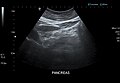
 Pancreas as seen on abdominal ultrasonography with Doppler.
 -
 Pancreas as seen on abdominal ultrasonography.
 -
 The pancreas and its surrounding structures
 -
 Duodenum and pancreas. Deep dissection.


External links[edit]
| Anatomy of the liver, pancreas and biliary tree | ||||||
|---|---|---|---|---|---|---|
|
Gray's Anatomy[edit]
- Gray's Anatomy Contents
- Gray's Anatomy Subject Index
- About Classic Gray's Anatomy
- Glossary of anatomy terms
Anatomy atlases (external)[edit]
[1] - Anatomy Atlases
| Human systems and organs | ||||||||||||||
|---|---|---|---|---|---|---|---|---|---|---|---|---|---|---|
|

Medical Disclaimer: WikiMD is for informational purposes only and is not a substitute for professional medical advice. Content may be inaccurate or outdated and should not be used for diagnosis or treatment. Always consult your healthcare provider for medical decisions. Verify information with trusted sources such as CDC.gov and NIH.gov. By using this site, you agree that WikiMD is not liable for any outcomes related to its content. See full disclaimer.
Credits:Most images are courtesy of Wikimedia commons, and templates, categories Wikipedia, licensed under CC BY SA or similar.
Translate page: - East Asian
中文,
日本,
한국어,
South Asian
हिन्दी,
தமிழ்,
తెలుగు,
Urdu,
ಕನ್ನಡ,
Southeast Asian
Indonesian,
Vietnamese,
Thai,
မြန်မာဘာသာ,
বাংলা
European
español,
Deutsch,
français,
Greek,
português do Brasil,
polski,
română,
русский,
Nederlands,
norsk,
svenska,
suomi,
Italian
Middle Eastern & African
عربى,
Turkish,
Persian,
Hebrew,
Afrikaans,
isiZulu,
Kiswahili,
Other
Bulgarian,
Hungarian,
Czech,
Swedish,
മലയാളം,
मराठी,
ਪੰਜਾਬੀ,
ગુજરાતી,
Portuguese,
Ukrainian
