The Facial Nerve
Anatomy > Gray's Anatomy of the Human Body > IX. Neurology > 5g. The Facial Nerve
Henry Gray (1821–1865). Anatomy of the Human Body. 1918.
The Facial Nerve[edit]
(N. Facialis; Seventh Nerve)
The facial nerve (Figs. 788, 790) consists of a motor and a sensory part, the latter being frequently described under the name of the nervus intermedius (pars intermedii of Wrisberg)(Fig. 788). The two parts emerge at the lower border of the pons in the recess between the olive and the inferior peduncle, the motor part being the more medial, immediately to the lateral side of the sensory part is the acoustic nerve.

The motor part supplies somatic motor fibers to the muscles of the face, scalp, and auricle, the Buccinator and Platysma, the Stapedius, the Stylohyoideus, and posterior belly of the Digastricus; it also contains some sympathetic motor fibers which constitute the vasodilator nerves of the submaxillary and sublingual glands, and are conveyed through the chorda tympani nerve.
These are preganglionic fibers of the sympathetic system and terminate in the submaxillary ganglion and small ganglia in the hilus of the submaxillary gland. From these ganglia postganglionic fibers are conveyed to these glands. The sensory part contains the fibers of taste for the anterior two-thirds of the tongue and a few somatic sensory fibers from the middle ear region. A few splanchnic sensory fibers are also present.
The motor root[edit]
The motor root arises from a nucleus which lies deeply in the reticular formation of the lower part of the pons. This nucleus is situated above the nucleus ambiguus, behind the superior olivary nucleus, and medial to the spinal tract of the trigeminal nerve.
From this origin the fibers pursue a curved course in the substance of the pons. They first pass backward and medialward toward the rhomboid fossa, and, reaching the posterior end of the nucleus of the abducent nerve, run upward close to the middle line beneath the colliculus fasciculus. At the anterior end of the nucleus of the abducent nerve they make a second bend, and run downward and forward through the pons to their point of emergence between the olive and the inferior peduncle.
The sensory root[edit]
The sensory root arises from the genicular ganglion, which is situated on the geniculum of the facial nerve in the facial canal, behind the hiatus of the canal. The cells of this ganglion are unipolar, and the single process divides in a T-shaped manner into central and peripheral branches. The central branches leave the trunk of the facial nerve in the internal acoustic meatus, and form the sensory root; the peripheral branches are continued into the chorda tympani and greater superficial petrosal nerves. Entering the brain at the lower border of the pons between the motor root and the acoustic nerve, the fibers of the sensory root pass into the substance of the medulla oblongata and end in the upper part of the terminal nucleus of the glossopharyngeal nerve and in the fasciculus solitarius.
FIG. 789– The course and connections of the facial nerve in the temporal bone. (Picture From the Classic Gray's Anatomy)
From their superficial attachments to the brain, the two roots of the facial nerve pass lateralward and forward with the acoustic nerve to the internal acoustic meatus. In the meatus the motor root lies in a groove on the upper and anterior surface of the acoustic nerve, the sensory root being placed between them.
At the bottom of the meatus, the facial nerve enters the facial canal, which it traverses to its termination at the stylomastoid foramen. It is at first directed lateralward between the cochlea and vestibule toward the medial wall of the tympanic cavity; it then bends suddenly backward and arches downward behind the tympanic cavity to the stylomastoid foramen.
The point where it changes its direction is named the geniculum it presents a reddish gangliform swelling, the genicular ganglion (ganglion geniculi; geniculate ganglion; nucleus of the sensory root of the nerve)(Fig. 789). On emerging from the stylomastoid foramen, the facial nerve runs forward in the substance of the parotid gland, crosses the external carotid artery, and divides behind the ramus of the mandible into branches, from which numerous offsets are distributed over the side of the head, face, and upper part of the neck, supplying the superficial muscles in these regions. The branches and their offsets unite to form the parotid plexus

Branches of Communication[edit]
The branches of communication of the facial nerve may be arranged as follows:
In the internal acoustic meatus……………… With the acoustic nerve.
At the genicular ganglion…………………… With the sphenopalatine ganglion by the greater superficial petrosal nerve.
With the otic ganglion by a branch which joins the lesser superficial petrosal nerve.
With the sympathetic on the middle meningeal artery.
In the facial canal…………………………… With the auricular branch of the vagus.
At its exit from the stylomastoid foramen…… With the glossopharyngeal. With the vagus. With the great auricular.
With the auriculotemporal.
Behind the ear……………………………… With the lesser occipital.
On the face………………………………… With the trigeminal.
In the neck………………………………… With the cutaneous cervical.
In the internal acoustic meatus some minute filaments pass from the facial to the acoustic nerve.
The greater superficial petrosal nerve (large superficial petrosal nerve) arises from the genicular ganglion, and consists chiefly of sensory branches which are distributed to the mucous membrane of the soft palate; but it probably contains a few motor fibers which form the motor root of the sphenopalatine ganglion.
It passes forward through the hiatus of the facial canal, and runs in a sulcus on the anterior surface of the petrous portion of the temporal bone beneath the semilunar ganglion, to the foramen lacerum.
It receives a twig from the tympanic plexus, and in the foramen is joined by the deep petrosal, from the sympathetic plexus on the internal carotid artery, to form the nerve of the pterygoid canal which passes forward through the pterygoid canal and ends in the sphenopalatine ganglion. The genicular ganglion is connected with the otic ganglion by a branch which joins the lesser superficial petrosal nerve, and also with the sympathetic filaments accompanying the middle meningeal artery.
According to Arnold, a twig passes back from the ganglion to the acoustic nerve. Just before the facial nerve emerges from the stylomastoid foramen, it generally receives a twig from the auricular branch of the vagus.
After its exit from the stylomastoid foramen, the facial nerve sends a twig to the glossopharyngeal, and communicates with the auricular branch of the vagus, with the great auricular nerve of the cervical plexus, with the auriculotemporal nerve in the parotid gland, and with the lesser occipital behind the ear; on the face with the terminal branches of the trigeminal, and in the neck with the cutaneous cervical nerve.
Branches of Distribution[edit]
The branches of distribution (Fig. 788) of the facial nerve may be thus arranged:
With the facial canal………………………….. Nerve to the Stapedius muscle.
At its exit from the stylomastoid foramen……… Posterior auricular.
On the face…………………………………… Temporal.
The Nerve to the Stapedius (n. stapedius; tympanic branch) arises opposite the pyramidal eminence (page 1042); it passes through a small canal in this eminence to reach the muscle.
The Chorda Tympani Nerve is given off from the facial as it passes downward behind the tympanic cavity, about 6 mm. from the stylomastoid foramen. It runs upward and forward in a canal, and enters the tympanic cavity, through an aperture (iter chordae posterius) on its posterior wall, close to the medial surface of the posterior border of the tympanic membrane and on a level with the upper end of the manubrium of the malleus.
It traverses the tympanic cavity, between the fibrous and mucous layers of the tympanic membrane, crosses the manubrium of the malleus, and emerges from the cavity through a foramen situated at the inner end of the petrotympanic fissure, and named the iter chordae anterius (canal of Huguier).
It then descends between the Pterygoideus externus and internus on the medial surface of the spina angularis of the sphenoid, which it sometimes grooves, and joins, at an acute angle, the posterior border of the lingual nerve. It receives a few efferent fibers from the motor root; these enter the submaxillary ganglion, and through it are distributed to the submaxillary and sublingual glands; the majority of its fibers are afferent, and are continued onward through the muscular substance of the tongue to the mucous membrane covering its anterior two-thirds; they constitute the nerve of taste for this portion of the tongue. Before uniting with the lingual nerve the chorda tympani is joined by a small branch from the otic ganglion.
The Posterior Auricular Nerve (n. auricularis posterior) arises close to the stylo-mastoid foramen, and runs upward in front of the mastoid process; here it is joined by a filament from the auricular branch of the vagus, and communicates with the posterior branch of the great auricular, and with the lesser occipital. As it ascends between the external acoustic meatus and mastoid process it divides into auricular and occipital branches.
The auricular branch supplies the Auricularis posterior and the intrinsic muscles on the cranial surface of the auricula.
The occipital branch the larger, passes backward along the superior nuchal line of the occipital bone, and supplies the Occipitalis.
The Digastric Branch (ramus digastricus) arises close to the stylomastoid foramen, and divides into several filaments, which supply the posterior belly of the Digastricus; one of these filaments joins the glossopharyngeal nerve.
The Stylohyoid Branch (ramus stylohyoideus) frequently arises in conjunction with the digastric branch; it is long and slender, and enters the Stylohyoideus about its middle.
The Temporal Branches (rami temporales) cross the zygomatic arch to the temporal region, supplying the Auriculares anterior and superior, and joining with the zygomaticotemporal branch of the maxillary, and with the auriculotemporal branch of the mandibular. The more anterior branches supply the Frontalis, the Orbicularis oculi, and the Corrugator, and join the supraorbital and lacrimal branches of the ophthalmic.
The Zygomatic Branches (rami zygomatici; malar branches) run across the zygomatic bone to the lateral angle of the orbit, where they supply the Orbicularis oculi, and join with filaments from the lacrimal nerve and the zygomaticofacial branch of the maxillary nerve.
The Buccal Branches (rami buccales; infraorbital branches), of larger size than the rest, pass horizontally forward to be distributed below the orbit and around the mouth. The superficial branches run beneath the skin and above the superficial muscles of the face, which they supply: some are distributed to the Procerus, joining at the medial angle of the orbit with the infratrochlear and nasociliary branches of the ophthalmic.
The deep branches pass beneath the Zygomaticus and the Quadratus labii superioris, supplying them and forming an infraorbital plexus with the infraorbital branch of the maxillary nerve. These branches also supply the small muscles of the nose. The lower deep branches supply the Buccinator and Orbicularis oris, and join with filaments of the buccinator branch of the mandibular nerve.
The Mandibular Branch (ramus marginalis mandibulae) passes forward beneath the Platysma and Triangularis, supplying the muscles of the lower lip and chin, and communicating with the mental branch of the inferior alveolar nerve.
The Cervical Branch (ramus colli) runs forward beneath the Platysma, and forms a series of arches across the side of the neck over the suprahyoid region. One branch descends to join the cervical cutaneous nerve from the cervical plexus; others supply the Platysma.
Facial sensation[edit]
In addition, the facial nerve receives taste sensations from the anterior two-thirds of the tongue via the chorda tympani. Taste sensation is sent to the gustatory portion (superior part) of the solitary nucleus. General sensation from the anterior two-thirds of tongue are supplied by afferent fibers of the third division of the fifth cranial nerve (V-3). These sensory (V-3) and taste (VII) fibers travel together as the lingual nerve briefly before the chorda tympani leaves the lingual nerve to enter the tympanic cavity (middle ear) via the petrotympanic fissure. It joins the rest of the facial nerve via the canaliculus for chorda tympani. The facial nerve then forms the geniculate ganglion, which contains the cell bodies of the taste fibers of chorda tympani and other taste and sensory pathways. From the geniculate ganglion, the taste fibers continue as the intermediate nerve which goes to the upper anterior quadrant of the fundus of the internal acoustic meatus along with the motor root of the facial nerve. The intermediate nerve reaches the posterior cranial fossa via the internal acoustic meatus before synapsing in the solitary nucleus.
The facial nerve also supplies a small amount of afferent innervation to the oropharynx below the palatine tonsil. There is also a small amount of cutaneous sensation carried by the nervus intermedius from the skin in and around the auricle (outer ear).
Other[edit]
The facial nerve also supplies parasympathetic fibers to the submandibular gland and sublingual glands via chorda tympani. Parasympathetic innervation serves to increase the flow of saliva from these glands. It also supplies parasympathetic innervation to the nasal mucosa and the lacrimal gland via the pterygopalatine ganglion. The parasympathetic fibers that travel in the facial nerve originate in the superior salivatory nucleus.
The facial nerve also functions as the efferent limb of the corneal reflex.
Functional components[edit]
The facial nerve carries axons of type GSA, general somatic afferent, to skin of the posterior ear.
The facial nerve also carries axons of type GVE, general visceral efferent, which innervate the sublingual, submandibular, and lacrimal glands, also mucosa of nasal cavity.
Axons of type SVE, special visceral efferent, innervate muscles of facial expression, stapedius, the posterior belly of digastric, and the stylohyoid.
The axons of type SVA, special visceral afferent, provide taste to the anterior two-thirds of tongue via chorda tympani.
Additional images[edit]
-
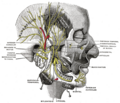
 Inferior view of the human brain, with the cranial nerves labelled.
 -
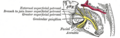
 Mandibular division of the trifacial nerve.
 -
 The course and connections of the facial nerve in the temporal bone.
 -
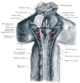 Upper part of medulla spinalis and hind- and mid-brains; posterior aspect, exposed in situ.
 -
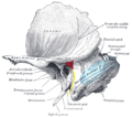 Left temporal bone showing surface markings for the tympanic antrum (red), transverse sinus (blue), and facial nerve (yellow).
 -
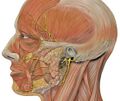 Head facial nerve branches
 -
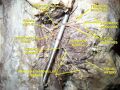 Facial nerve. Deep dissection.


External links[edit]
| The cranial nerves | ||||||||||
|---|---|---|---|---|---|---|---|---|---|---|
|
Gray's Anatomy[edit]
- Gray's Anatomy Contents
- Gray's Anatomy Subject Index
- About Classic Gray's Anatomy
- Glossary of anatomy terms
Anatomy atlases (external)[edit]
[1] - Anatomy Atlases
| Human systems and organs | ||||||||||||||
|---|---|---|---|---|---|---|---|---|---|---|---|---|---|---|
|

Medical Disclaimer: WikiMD is for informational purposes only and is not a substitute for professional medical advice. Content may be inaccurate or outdated and should not be used for diagnosis or treatment. Always consult your healthcare provider for medical decisions. Verify information with trusted sources such as CDC.gov and NIH.gov. By using this site, you agree that WikiMD is not liable for any outcomes related to its content. See full disclaimer.
Credits:Most images are courtesy of Wikimedia commons, and templates, categories Wikipedia, licensed under CC BY SA or similar.
Translate page: - East Asian
中文,
日本,
한국어,
South Asian
हिन्दी,
தமிழ்,
తెలుగు,
Urdu,
ಕನ್ನಡ,
Southeast Asian
Indonesian,
Vietnamese,
Thai,
မြန်မာဘာသာ,
বাংলা
European
español,
Deutsch,
français,
Greek,
português do Brasil,
polski,
română,
русский,
Nederlands,
norsk,
svenska,
suomi,
Italian
Middle Eastern & African
عربى,
Turkish,
Persian,
Hebrew,
Afrikaans,
isiZulu,
Kiswahili,
Other
Bulgarian,
Hungarian,
Czech,
Swedish,
മലയാളം,
मराठी,
ਪੰਜਾਬੀ,
ગુજરાતી,
Portuguese,
Ukrainian
