The Spermatozoön
Anatomy>Gray's Anatomy of the Human Body > I. Embryology > 3.The Spermatozoon Henry Gray (1821–1865). Anatomy of the Human Body. 1918. A spermatozoon, alternate spelling spermatozoön; plural spermatozoa; from Ancient Greek: σπέρμα
("seed") and ("living being")) is a motile sperm cell, or moving form of the haploid cell that is the male gamete. A spermatozoon joins an ovum to form a zygote. (A zygote is a single cell, with a complete set of chromosomes, that normally develops into an embryo.)
Sperm cells contribute approximately half of the nuclear genetic information to the diploid offspring (excluding, in most cases, mitochondrial DNA). In mammals, the sex of the offspring is determined by the sperm cell: a spermatozoon bearing a X chromosome will lead to a female (XX) offspring, while one bearing a Y chromosome will lead to a male (XY) offspring. Sperm cells were first observed in Antonie van Leeuwenhoek's laboratory in 1677.
Humans[edit]
The human sperm cell is the reproductive cell in males and will only survive in warm environments; once it leaves the male body the sperm's survival likelihood is reduced and it may die, thereby decreasing the total sperm quality. Sperm cells come in two types, "female" and "male". Sperm cells that give rise to female (XX) offspring after fertilization differ in that they carry an X-chromosome, while sperm cells that give rise to male (XY) offspring carry a Y-chromosome.
A human sperm cell consists of a flat, disc shaped head 5.1 µm by 3.1 µm and a tail 50 µm long. The tail flagellates, which propels the sperm cell (at about 1–3 mm/minute in humans) by whipping in an elliptical cone. Sperm have an olfactory guidance mechanism, and after reaching the Fallopian tubes, must undergo a period of capacitation before penetration of the ovum.
Head[edit]
It has a compact nucleus with only chromatic substance and is surrounded by only a thin rim of cytoplasm. Above the nucleus lies a cap-like structure called the acrosome, formed by modification of the Golgi body, which secretes the enzyme spermlysin (hyaluronidase, corona-penetrating enzyme, zona eyesin, or aerosin), that are necessary for fertilization. The acrosomal region experiment the acrosomal reaction, it consists in the fusion of the sperm plasma membrane with the outer acrosomal membrane. On the surface of the head lies a decapacitating substance which is removed before fertilisation.
Neck[edit]
It is the smallest part (0.03 ×10⁻⁶ m), and has a proximal centriole parallel to the base of the nucleus and distal centriole perpendicular to the previous one. The proximal centriole is present also in the mature spermatozoon; the distal centriole disappears after axoneme assembly. The proximal centriole enters into the egg during fertilisation and starts the first cleavage division of the egg, which has no centriole. The distal centriole gives rise to the axial filament which forms the tail and has a (9+2) arrangement. A transitory membrane called the Manchette lies in the middle piece.
Middle piece[edit]
It has 10–14 spirals of mitochondria surrounding the axial filament in the cytoplasm. It provides motility, and hence is called the powerhouse of the sperm. It also has a ring centriole (annulus) that form a diffusion barrier between the middle piece and the principal piece and serve as a stabilizing structure for tail rigidity.
Tail[edit]
It is the longest part (50×10−6 m), having an axial filament surrounded by cytoplasm and plasma membrane, but at the posterior end the axial filament is naked. It is push mechanism.
Semen has an alkaline nature and the spermatozoa do not reach full motility (hypermotility) until they reach the vagina, where the alkaline pH is neutralized by acidic vaginal fluids. This gradual process takes 20–30 minutes. During this period, fibrinogen from the seminal vesicles forms a clot, securing and protecting the sperm. Just as they become hypermotile, fibrinolysin from the prostate gland dissolves the clot, allowing the sperm to progress optimally.
The spermatozoon is characterized by a minimum of cytoplasm and the most densely packed DNA known in eukaryotes. Compared to mitotic chromosomes in somatic cells, sperm DNA is at least sixfold more highly condensed.
The specimen contributes with DNA/chromatin, a centriole, and perhaps also an oocyte-activating factor (OAF).
-
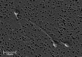 Electron micrograph of human spermatozoa magnified 3140 times.
 -
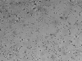 Sperm cells in the urine sample of a 45-year-old male patient who is being followed with the diagnosis of benign prostate hyperplasia.
 -
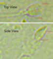 Dimensions of the human sperm head measured from a 39 year-old healthy subject.

_-_Spermler_(idrar)_-_01.png)

The human spermatozoon contains at least 7500 different proteins.
Human sperm genetics has been associated with human evolution, per a 2020 study.

Spermatozoa production in mammals[edit]
Spermatozoa are produced in the seminiferous tubules of the testes in a process called spermatogenesis. Round cells called spermatogonia divide and differentiate eventually to become spermatozoa. During copulation the cloaca or vagina gets inseminated, and then the spermatozoa move through chemotaxis to the ovum inside a Fallopian tube or the uterus.
Artificial storage[edit]
Spermatozoa can be stored in diluents such as the Illini Variable Temperature (IVT) diluent, which have been reported to be able to preserve high fertility of spermatozoa for over seven days.
Semen cryopreservation can be used for far longer storage durations. For human spermatozoa, the longest reported successful storage with this method is 21 years.
External links[edit]
| Sex | ||||||
|---|---|---|---|---|---|---|
* Category
|
| Antonie van Leeuwenhoek | ||||||||||||||
|---|---|---|---|---|---|---|---|---|---|---|---|---|---|---|
|
Gray's Anatomy[edit]
- Gray's Anatomy Contents
- Gray's Anatomy Subject Index
- About Classic Gray's Anatomy
- Glossary of anatomy terms
Anatomy atlases (external)[edit]
[1] - Anatomy Atlases
| Human systems and organs | ||||||||||||||
|---|---|---|---|---|---|---|---|---|---|---|---|---|---|---|
|

Medical Disclaimer: WikiMD is for informational purposes only and is not a substitute for professional medical advice. Content may be inaccurate or outdated and should not be used for diagnosis or treatment. Always consult your healthcare provider for medical decisions. Verify information with trusted sources such as CDC.gov and NIH.gov. By using this site, you agree that WikiMD is not liable for any outcomes related to its content. See full disclaimer.
Credits:Most images are courtesy of Wikimedia commons, and templates, categories Wikipedia, licensed under CC BY SA or similar.
Translate page: - East Asian
中文,
日本,
한국어,
South Asian
हिन्दी,
தமிழ்,
తెలుగు,
Urdu,
ಕನ್ನಡ,
Southeast Asian
Indonesian,
Vietnamese,
Thai,
မြန်မာဘာသာ,
বাংলা
European
español,
Deutsch,
français,
Greek,
português do Brasil,
polski,
română,
русский,
Nederlands,
norsk,
svenska,
suomi,
Italian
Middle Eastern & African
عربى,
Turkish,
Persian,
Hebrew,
Afrikaans,
isiZulu,
Kiswahili,
Other
Bulgarian,
Hungarian,
Czech,
Swedish,
മലയാളം,
मराठी,
ਪੰਜਾਬੀ,
ગુજરાતી,
Portuguese,
Ukrainian
