The Uterine Tube
Anatomy > Gray's Anatomy of the Human Body > XI. Splanchnology > 3d. 2. The Uterine Tube
Henry Gray (1821–1865). Anatomy of the Human Body. 1918.
The Uterine Tube[edit]
(Tuba Uterina [Fallopii]; Fallopian Tube; Oviduct)
The uterine tubes (Figs. 1161, 1165) convey the ova from the ovaries to the cavity of the uterus. They are two in number, one on either side, situated in the upper margin of the broad ligament, and extending from the superior angle of the uterus to the side of the pelvis.
Each tube is about 10 cm. long, and is described as consisting of three portions:
(1) the isthmus or medial constricted third;
(2) the ampulla or intermediate dilated portion, which curves over the ovary; and
(3) the infundibulum with its abdominal ostium surrounded by fimbriae one of which, the ovarian fimbria is attached to the ovary. The uterine tube is directed lateralward as far as the uterine pole of the ovary, and then ascends along the mesovarian border of the ovary to the tubal pole, over which it arches; finally it turns downward and ends in relation to the free border and medial surface of the ovary. The uterine opening is minute, and will only admit a fine bristle; the abdominal opening is somewhat larger. In connection with the fimbriae of the uterine tube, or with the broad ligament close to them, there are frequently one or more small pedunculated vesicles. These are termed the appendices vesiculosae (hydatids of Morgagni).

Structure[edit]
The uterine tube consists of three coats: serous, muscular and mucous
The external or serous coat is peritoneal.
The middle or muscular coat consists of an external longitudinal and an internal circular layer of non-striped muscular fibers continuous with those of the uterus.
The internal or mucous coat is continuous with the mucous lining of the uterus, and, at the abdominal ostium of the tube, with the peritoneum. It is thrown into longitudinal folds, which in the ampulla are much more extensive than in the isthmus. The lining epithelium is columnar and ciliated. This form of epithelium is also found on the inner surface of the fimbriae. while on the outer or serous surfaces of these processes the epithelium gradually merges into the endothelium of the peritoneum.
Fertilization of the ovum is believed (page 44) to occur in the tube, and the fertilized ovum is then normally passed on into the uterus; the ovum, however, may adhere to and undergo development in the uterine tube, giving rise to the commonest variety of ectopic gestation In such cases the amnion and chorion are formed, but a true decidua is never present; and the gestation usually ends by extrusion of the ovum through the abdominal ostium, although it is not uncommon for the tube to rupture into the peritoneal cavity, this being accompanied by severe hemorrhage, and needing surgical interference.
Function[edit]
Fertilization[edit]
The fallopian tube allows passage of an egg from the ovary to the uterus. When an oocyte is developing in an ovary, it is surrounded by a spherical collection of cells known as an ovarian follicle. Just before ovulation, the primary oocyte completes meiosis I to form the first polar body and a secondary oocyte which is arrested in metaphase of meiosis II.
At the time of ovulation in the menstrual cycle, the secondary oocyte is released from the ovary. The follicle and the ovary's wall rupture, allowing the secondary oocyte to escape. The secondary oocyte is caught by the fimbriated end of the Fallopian tube and travels to the ampulla. Here, the egg is able to become fertilised with sperm. The ampulla is typically where the sperm are met and fertilization occurs; meiosis II is promptly completed. After fertilisation, the ovum is now called a zygote and travels towards the uterus with the aid of the hair-like cilia and the activity of the muscle of the Fallopian tube. The early embryo requires critical development in the fallopian tube.[1] After about five days the new embryo enters the uterine cavity and on about the sixth day implants on the wall of the uterus.
The release of an oocyte does not alternate between the two ovaries and seems to be random. After removal of an ovary, the remaining one produces an egg every month.[2]
Additional images[edit]
-
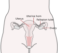 Image showing the position of the Fallopian tubes between the ovaries and the uterus.
 -
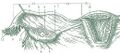 Numbered image showing parts of the Fallopian tubes and surrounding structures. 1: Ovary
2: Medial surface
3: Lateral surface
4: Free border
5: Mesovarial margin
6: Tubal extremity
7: Uterine extremity
8: Fallopian tube
9: Distal opening of the Fallopian tube
10: Infundibulum of the Fallopian tube
11: Fimbriae of the Fallopian tube
12: Ovarian fimbria
13: Ampulla of the Fallopian tube
14: Isthmus of the Fallopian tube
15: Uterine part of the Fallopian tube
16: Proximal opening of the Fallopian tube -
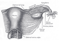 Image showing the right Fallopian tube (here labelled the uterine tube) seen from behind. The uterus, ovaries and right broad ligament are labelled.
 -
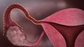 Unlabelled image showing the right Fallopian tube
 -
Isthmus of the Fallopian tube seen arising from the uterus in a cadaveric specimen.
.svg)


External links[edit]
- Histology image: 18501loa – Histology Learning System at Boston University
| Female reproductive system | ||||||||||||||||||||
|---|---|---|---|---|---|---|---|---|---|---|---|---|---|---|---|---|---|---|---|---|
|
Gray's Anatomy[edit]
- Gray's Anatomy Contents
- Gray's Anatomy Subject Index
- About Classic Gray's Anatomy
- Glossary of anatomy terms
Anatomy atlases (external)[edit]
[1] - Anatomy Atlases
| Human systems and organs | ||||||||||||||
|---|---|---|---|---|---|---|---|---|---|---|---|---|---|---|
|

Medical Disclaimer: WikiMD is for informational purposes only and is not a substitute for professional medical advice. Content may be inaccurate or outdated and should not be used for diagnosis or treatment. Always consult your healthcare provider for medical decisions. Verify information with trusted sources such as CDC.gov and NIH.gov. By using this site, you agree that WikiMD is not liable for any outcomes related to its content. See full disclaimer.
Credits:Most images are courtesy of Wikimedia commons, and templates, categories Wikipedia, licensed under CC BY SA or similar.
Translate page: - East Asian
中文,
日本,
한국어,
South Asian
हिन्दी,
தமிழ்,
తెలుగు,
Urdu,
ಕನ್ನಡ,
Southeast Asian
Indonesian,
Vietnamese,
Thai,
မြန်မာဘာသာ,
বাংলা
European
español,
Deutsch,
français,
Greek,
português do Brasil,
polski,
română,
русский,
Nederlands,
norsk,
svenska,
suomi,
Italian
Middle Eastern & African
عربى,
Turkish,
Persian,
Hebrew,
Afrikaans,
isiZulu,
Kiswahili,
Other
Bulgarian,
Hungarian,
Czech,
Swedish,
മലയാളം,
मराठी,
ਪੰਜਾਬੀ,
ગુજરાતી,
Portuguese,
Ukrainian
