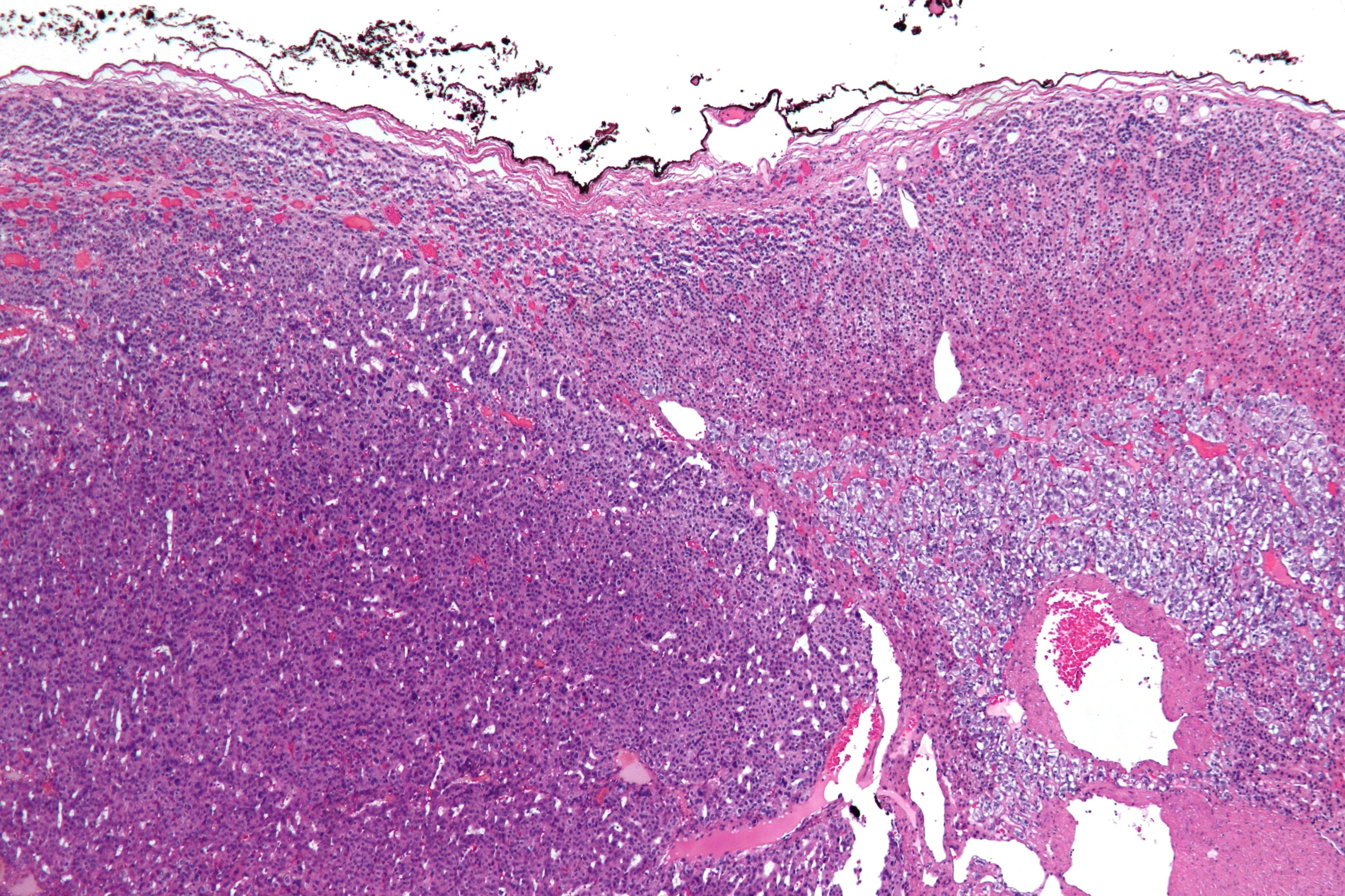
Adrenocortical carcinoma
(Redirected from Adrenal carcinoma)
| Adrenocortical carcinoma | |
|---|---|

| |
| Synonyms | Adrenal cortical carcinoma, ACC |
| Pronounce | N/A |
| Specialty | N/A |
| Symptoms | Abdominal pain, weight loss, virilization, Cushing's syndrome |
| Complications | Metastasis, hormonal imbalance |
| Onset | Most common in adults aged 40-50 |
| Duration | Chronic |
| Types | Functioning, non-functioning |
| Causes | Unknown |
| Risks | Li-Fraumeni syndrome, Beckwith-Wiedemann syndrome, Multiple endocrine neoplasia |
| Diagnosis | CT scan, MRI, biopsy |
| Differential diagnosis | Adrenal adenoma, pheochromocytoma, renal cell carcinoma |
| Prevention | N/A |
| Treatment | Surgery, mitotane, chemotherapy, radiation therapy |
| Medication | N/A |
| Prognosis | Generally poor, depends on stage |
| Frequency | Rare, 1-2 per million per year |
| Deaths | High mortality rate |
Other Names: ACC
Adrenocortical carcinoma is a rare disease in which malignant (cancer) cells form in the outer layer of the adrenal gland. Adrenocortical carcinoma is also called cancer of the adrenal cortex.
A tumor of the adrenal cortex may be functioning (makes more hormones than normal) or nonfunctioning (does not make more hormones than normal). Most adrenocortical tumors are functioning. The hormones made by functioning tumors may cause certain signs or symptoms of disease. The adrenal medulla makes hormones that help the body react to stress. Cancer that forms in the adrenal medulla is called pheochromocytoma.
Cause[edit | edit source]
There are a number of genes that have changes (mutations) that can cause an adrenocortical carcinoma, including TP53 and IGF2.
Inheritance[edit | edit source]
There have been reports of both autosomal dominant inheritance and autosomal recessive inheritance.
Riskfactors[edit | edit source]
Risk factors for adrenocortical carcinoma include having the following hereditary diseases:
- Li-Fraumeni syndrome.
- Beckwith-Wiedemann syndrome.
- Carney complex.
Symptoms[edit | edit source]
Symptoms of adrenocortical carcinoma include pain in the abdomen. These and other signs and symptoms may be caused by adrenocortical carcinoma:
- A lump in the abdomen.
- Pain the abdomen or back.
- A feeling of fullness in the abdomen.
A nonfunctioning adrenocortical tumor may not cause signs or symptoms in the early stages.
A functioning adrenocortical tumor makes too much of one of the following hormones:
- Cortisol.
- Aldosterone.
- Testosterone.
- Estrogen.
Too much cortisol may cause:
- Weight gain in the face, neck, and trunk of the body and thin arms and legs.
- Growth of fine hair on the face, upper back, or arms.
- A round, red, full face.
- A lump of fat on the back of the neck.
- A deepening of the voice and swelling of the sex organs or breasts in both males and females.
- Muscle weakness.
- High blood sugar.
- High blood pressure.
Too much aldosterone may cause:
- High blood pressure.
- Muscle weakness or cramps.
- Frequent urination.
- Feeling thirsty.
Too much testosterone (in women) may cause:
- Growth of fine hair on the face, upper back, or arms.
- Acne.
- Balding.
- A deepening of the voice.
- No menstrual periods.
Men who make too much testosterone do not usually have signs or symptoms.
Too much estrogen (in women) may cause:
- Irregular menstrual periods in women who have not gone through menopause.
- Vaginal bleeding in women who have gone through menopause.
- Weight gain.
Too much estrogen (in men) may cause:
- Growth of breast tissue.
- Lower sex drive.
- Impotence.
Diagnosis[edit | edit source]
The tests and procedures used to diagnose adrenocortical carcinoma depend on the patient's signs and symptoms. The following tests and procedures may be used:
Physical exam and history: An exam of the body to check general signs of health, including checking for signs of disease, such as lumps or anything else that seems unusual. A history of the patient’s health habits and past illnesses and treatments will also be taken. Twenty-four-hour urine test: A test in which urine is collected for 24 hours to measure the amounts of cortisol or 17-ketosteroids. A higher than normal amount of these in the urine may be a sign of disease in the adrenal cortex.
Low-dose dexamethasone suppression test: A test in which one or more small doses of dexamethasone are given. The level of cortisol is checked from a sample of blood or from urine that is collected for three days. This test is done to check if the adrenal gland is making too much cortisol.
High-dose dexamethasone suppression test: A test in which one or more high doses of dexamethasone are given. The level of cortisol is checked from a sample of blood or from urine that is collected for three days. This test is done to check if the adrenal gland is making too much cortisol or if the pituitary gland is telling the adrenal glands to make too much cortisol.
Blood chemistry study: A procedure in which a blood sample is checked to measure the amounts of certain substances, such as potassium or sodium, released into the blood by organs and tissues in the body. An unusual (higher or lower than normal) amount of a substance can be a sign of disease.
CT scan (CAT scan): A procedure that makes a series of detailed pictures of areas inside the body, taken from different angles. The pictures are made by a computer linked to an x-ray machine. A dye may be injected into a vein or swallowed to help the organs or tissues show up more clearly. This procedure is also called computed tomography, computerized tomography, or computerized axial tomography.
MRI (magnetic resonance imaging): A procedure that uses a magnet, radio waves, and a computer to make a series of detailed pictures of areas inside the body. This procedure is also called nuclear magnetic resonance imaging (NMRI). An MRI of the abdomen is done to diagnose adrenocortical carcinoma.
Adrenal angiography: A procedure to look at the arteries and the flow of blood near the adrenal glands. A contrast dye is injected into the adrenal arteries. As the dye moves through the arteries, a series of x-rays are taken to see if any arteries are blocked.
Adrenal venography: A procedure to look at the adrenal veins and the flow of blood near the adrenal glands. A contrast dye is injected into an adrenal vein. As the contrast dye moves through the veins, a series of x-rays are taken to see if any veins are blocked. A catheter (very thin tube) may be inserted into the vein to take a blood sample, which is checked for abnormal hormone levels.
PET scan (positron emission tomography scan): A procedure to find malignant tumor cells in the body. A small amount of radioactive glucose (sugar) is injected into a vein. The PET scanner rotates around the body and makes a picture of where glucose is being used in the body. Malignant tumor cells show up brighter in the picture because they are more active and take up more glucose than normal cells do.
MIBG scan: A very small amount of radioactive material called MIBG is injected into a vein and travels through the bloodstream. Adrenal gland cells take up the radioactive material and are detected by a device that measures radiation. This scan is done to tell the difference between adrenocortical carcinoma and pheochromocytoma.
Biopsy: The removal of cells or tissues so they can be viewed under a microscope by a pathologist to check for signs of cancer. The sample may be taken using a thin needle, called a fine-needle aspiration (FNA) biopsy or a wider needle, called a core biopsy.
Treatment[edit | edit source]
Treatment options include surgical removal of the tumor, which is important to achieve a good long-term outlook. Chemotherapy, specifically a drug called mitotane, can be used to try to remove any remaining cancer after surgery. Surgery Surgery to remove the adrenal gland (adrenalectomy) is often used to treat adrenocortical carcinoma. Sometimes surgery is done to remove the nearby lymph nodes and other tissue where the cancer has spread.
Radiation therapy[edit | edit source]
Radiation therapy is a cancer treatment that uses high-energy x-rays or other types of radiation to kill cancer cells or keep them from growing. There are two types of radiation therapy:
External radiation therapy uses a machine outside the body to send radiation toward the cancer. Internal radiation therapy uses a radioactive substance sealed in needles, seeds, wires, or catheters that are placed directly into or near the cancer. The way the radiation therapy is given depends on the type and stage of the cancer being treated. External radiation therapy is used to treat adrenocortical carcinoma.
Chemotherapy Chemotherapy is a cancer treatment that uses drugs to stop the growth of cancer cells, either by killing the cells or by stopping them from dividing. When chemotherapy is taken by mouth or injected into a vein or muscle, the drugs enter the bloodstream and can reach cancer cells throughout the body (systemic chemotherapy). When chemotherapy is placed directly into the cerebrospinal fluid, an organ, or a body cavity such as the abdomen, the drugs mainly affect cancer cells in those areas (regional chemotherapy). Combination chemotherapy is treatment using more than one anticancer drug. The way the chemotherapy is given depends on the type and stage of the cancer being treated.
Prognosis[edit | edit source]
Adrenocortical carcinoma may be cured if treated at an early stage.
| Glandular and epithelial cancer | ||||||||||||||||||||||||
|---|---|---|---|---|---|---|---|---|---|---|---|---|---|---|---|---|---|---|---|---|---|---|---|---|
|
| Tumours of endocrine glands | ||||||||||
|---|---|---|---|---|---|---|---|---|---|---|
|
NIH genetic and rare disease info[edit source]
Adrenocortical carcinoma is a rare disease.
| Rare and genetic diseases | ||||||
|---|---|---|---|---|---|---|
|
Rare diseases - Adrenocortical carcinoma
|
Search WikiMD
Ad.Tired of being Overweight? Try W8MD's physician weight loss program.
Semaglutide (Ozempic / Wegovy and Tirzepatide (Mounjaro / Zepbound) available.
Advertise on WikiMD
|
WikiMD's Wellness Encyclopedia |
| Let Food Be Thy Medicine Medicine Thy Food - Hippocrates |
Translate this page: - East Asian
中文,
日本,
한국어,
South Asian
हिन्दी,
தமிழ்,
తెలుగు,
Urdu,
ಕನ್ನಡ,
Southeast Asian
Indonesian,
Vietnamese,
Thai,
မြန်မာဘာသာ,
বাংলা
European
español,
Deutsch,
français,
Greek,
português do Brasil,
polski,
română,
русский,
Nederlands,
norsk,
svenska,
suomi,
Italian
Middle Eastern & African
عربى,
Turkish,
Persian,
Hebrew,
Afrikaans,
isiZulu,
Kiswahili,
Other
Bulgarian,
Hungarian,
Czech,
Swedish,
മലയാളം,
मराठी,
ਪੰਜਾਬੀ,
ગુજરાતી,
Portuguese,
Ukrainian
Medical Disclaimer: WikiMD is not a substitute for professional medical advice. The information on WikiMD is provided as an information resource only, may be incorrect, outdated or misleading, and is not to be used or relied on for any diagnostic or treatment purposes. Please consult your health care provider before making any healthcare decisions or for guidance about a specific medical condition. WikiMD expressly disclaims responsibility, and shall have no liability, for any damages, loss, injury, or liability whatsoever suffered as a result of your reliance on the information contained in this site. By visiting this site you agree to the foregoing terms and conditions, which may from time to time be changed or supplemented by WikiMD. If you do not agree to the foregoing terms and conditions, you should not enter or use this site. See full disclaimer.
Credits:Most images are courtesy of Wikimedia commons, and templates, categories Wikipedia, licensed under CC BY SA or similar.
Contributors: Deepika vegiraju, Prab R. Tumpati, MD


