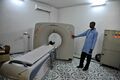
Mammography
.jpg)
- Mammography is a technique for the detection of breast cancer. In this procedure, several X-ray views are taken of one or both breasts and the radiographs are examined for the presence of a lesion.
- When a lesion is detected, the radiologist often can determine quite accurately whether it is malignant or benign.
- Mammography is important because very small, early cancers can be diagnosed with this technique before they are large enough to palpate. Mammograms of the opposite breast should be recorded as well as those of the involved breast. The findings of a mammographic examination will be reported on an x-ray report.
How does it work?[edit]
During a mammogram, a patient’s breast is placed on a flat support plate and compressed with a parallel plate called a paddle. An x-ray machine produces a small burst of x-rays that pass through the breast to a detector located on the opposite side. The detector can be either a photographic film plate, which captures the x-ray image on film, or a solid-state detector, which transmits electronic signals to a computer to form a digital image. The images produced are called mammograms.
On a film mammogram, low density tissues, such as fat, appear translucent (i.e. darker shades of gray approaching the black background)., whereas areas of dense tissue, such as connective and glandular tissue or tumors, appear whiter on a gray background. In a standard mammogram, both a top and a side view are taken of each breast, although extra views may be taken if the physician is concerned about a suspicious area of the breast.
What will the results look like?[edit]
A radiologist will carefully examine a mammogram to search for high density regions or areas of unusual configuration that look different from normal tissue. These areas could represent many different types of abnormalities, including cancerous tumors, non-cancerous masses called benign tumors, fibroadenomas, or complex cysts. Radiologists look at the size, shape, and contrast of an abnormal region, as well as the appearance of the edges or margins of such an area, all of which can indicate the possibility of malignancy (i.e. cancer). They also look for tiny bits of calcium, called microcalcifications, which show up as very bright specks on a mammogram. While usually benign, sites of microcalcifications may occasionally signal the presence of a specific type of cancer. If a mammogram shows one or more suspicious regions that are not definitive for cancer, the radiologist may order additional mammogram views, with or without additional magnification or compression, or they may order a biopsy. Another alternative may be referral for another type of non-invasive imaging study.

Why does the breast need to be compressed?[edit]
Compression holds the breast in place to minimize blurring of the x-ray image that can be caused by patient motion. Also, compression evens out the shape of the breast so that the x-rays can travel through a shorter path to reach the detector. This reduces the radiation dose and improves the quality of the x-ray image. Finally, compression allows all the tissues to be visualized in a single plane so that small abnormalities are less likely to be obscured by overlying breast tissue.
What is digital mammography?[edit]
A digital mammogram uses the same x-ray technology as conventional mammograms, but instead of using film, solid-state detectors are used to record the x-ray pattern passing through the breast. These detectors convert the x-rays that pass through them into electronic signals that are sent to a computer. The computer then converts these electronic signals into images that can be displayed on a monitor and also stored for later use. Several advantages of using digital mammography over film mammography include: the ability to manipulate the image contrast for better clarity, the ability to use computer-aided detection of abnormalities, and the ability to easily transmit digital files to other experts for a second opinion. In addition, digital mammograms may decrease the need for the re-takes, which are common with film mammography due to incorrect exposure techniques or problems with film development. As a result, digital mammography can lead to lower x-ray exposures. To date, there is no evidence that digital mammography is better that film mammography for reducing a woman’s risk of dying from breast cancer, however, digital screening may be more accurate for finding cancers in younger women or women with dense breasts. Read more on the National Cancer Institute’s webpage on mammograms.
What is tomosynthesis (3D mammography)?[edit]
Digital Breast Tomosynthesis, also known as 3D mammography, is an FDA-approved method for breast cancer screening in which x-rays of the breast are taken at different angles to generate thin cross-sections. The 3D representation of the breast is similar to the 3d images created by standard CT technology. Tomosynthesis differs from CT technology in that significantly fewer x-ray beams are projected through the breast than with CT and the x-ray exposure to the rest of the chest is dramatically reduced. Hence, the radiation dose delivered to the breast by tomosynthesis is similar to that delivered 2D mammography. While tomosynthesis uses very low-dose x-rays, it is currently most often used in addition to 2D mammography, making the total radiation dose higher than standard mammography. Early evaluations of 3D mammography suggest an improved detection of breast cancers than seen with 2D mammography, but extensive large-scale comparisons of tomosynthesis with 2D mammography in randomized studies are still in process. Therefore, researchers do not know with full certainty whether 3D mammography is better or worse than standard mammography at avoiding false-positive results and identifying early cancers in all types of patients.
What are the limits of mammography?[edit]
For certain types of breasts, mammograms can be difficult to interpret. This is because there is a wide variation in breast tissue density among women. Denser breasts are more difficult to image, and more difficult to assess for tumor diagnosis. For this and other reasons, the sensitivity of mammography in detecting cancer can vary over a wide range.
For many difficult cases, x-ray mammography alone may not be sufficiently sensitive or accurate in detecting cancer, so additional imaging technologies, such as ultrasound or magnetic resonance imaging (MRI) may also be used to increase the sensitivity of the exam. Recently, studies have shown that a type of nuclear medicinecalled molecular breast imaging (MBI) may be an effective and less expensive alternative to MRI for clarifying test results in patients with dense breasts. During MBI, patients are given an injection of radioactive molecules that are selectively taken up by cancer cells. Special cameras that detect radioactivity are then used to reveal these cancer cells in the breast tissue.
Are there risks?[edit]
Because mammography uses x-rays to produce images of the breast, patients are exposed to a small amount of ionizing radiation. For most women, the benefits of regular mammograms outweigh the risks posed by this amount of radiation. The risk associated with this dose appears to be greater among younger women (under age 40). However, in some cases, the benefits of using mammography to detect breast cancer under age 40 may outweigh the risks of radiation exposure. For example, a mammogram may reveal that a suspicious mass is benign and, therefore, doesn’t need to be treated. Additionally, if a tumor is malignant and is caught early by mammogram, a surgeon may be able to remove it before it spreads and requires more aggressive treatment such as chemotherapy.
When should I get a mammogram?[edit]
Several organizations and professional societies have developed guidelines for mammography screening. You can read more about these recommendations on their websites below. All recommend that women talk with their doctor about the benefits and potential harms of mammography, when to start screening, and how often to be screened.

What is the best method of detecting breast cancer as early as possible?[edit]
A high-quality mammogram plus a clinical breast exam, an exam done by your doctor, is the most effective way to detect breast cancer early. Finding breast cancer early greatly improves a woman's chances for successful treatment.
Like any test, mammograms have both benefits and limitations. For example, some cancers can't be found by a mammogram, but they may be found in a clinical breast exam.
Checking your own breasts for lumps or other changes is called a breast self-exam (BSE). Studies so far have not shown that BSE alone helps reduce the number of deaths from breast cancer. BSE should not take the place of routine clinical breast exams and mammograms.
If you choose to do BSE, remember that breast changes can occur because of pregnancy, aging, menopause, menstrual cycles, or from taking birth control pills or other hormones. It is normal for breasts to feel a little lumpy and uneven. Also, it is common for breasts to be swollen and tender right before or during a menstrual period. If you notice any unusual changes in your breasts, contact your doctor.

What are the steps in doing mammography?[edit]
- You stand in front of a special x-ray machine.
- The person who takes the x-rays, called a radiologic technician, places your breasts, one at a time, between an x-ray plate and a plastic plate.
- These plates are attached to the x-ray machine and compress the breasts to flatten them.
- This spreads the breast tissue out to obtain a clearer picture. You will feel pressure on your breast for a few seconds.
- It may cause you some discomfort; you might feel squeezed or pinched.
- This feeling only lasts for a few seconds, and the flatter your breast, the better the picture.
- Most often, two pictures are taken of each breast — one from the side and one from above.
- A screening mammogram takes about 20 minutes from start to finish.
How often should I get a mammogram?[edit]
The United States Preventive Services Task Force (USPSTF) recommends:
- Women ages 50 to 74 years should get a mammogram every 2 years.
- Women younger than age 50 should talk to a doctor about when to start and how often to have a mammogram.
What can mammograms show?[edit]
The radiologist will look at your x-rays for breast changes that do not look normal and for differences in each breast. He or she will compare your past mammograms with your most recent one to check for changes. The doctor will also look for lumps and calcifications.
- Breast Lump or mass. The size, shape, and edges of a lump sometimes can give doctors information about whether or not it may be cancer. On a mammogram, a growth that is benign often looks smooth and round with a clear, defined edge. Breast cancer often has a jagged outline and an irregular shape.
- Breast calcification. A calcification is a deposit of the mineral calcium in the breast tissue. Calcifications appear as small white spots on a mammogram. There are two types:
- Macrocalcifications are large calcium deposits often caused by aging. These usually are not a sign of cancer.
- Microcalcifications are tiny specks of calcium that may be found in an area of rapidly dividing cells.
If calcifications are grouped together in a certain way, it may be a sign of cancer. Depending on how many calcium specks you have, how big they are, and what they look like, your doctor may suggest that you have other tests. Calcium in the diet does not create calcium deposits, or calcifications, in the breast.
What if my screening mammogram shows a problem?[edit]
If you have a screening test result that suggests cancer, your doctor must find out whether it is due to cancer or to some other cause. Your doctor may ask about your personal and family medical history. You may have a physical exam. Your doctor also may order some of these tests:
- Diagnostic mammogram, to focus on a specific area of the breast
- Ultrasound, an imaging test that uses sound waves to create a picture of your breast. The pictures may show whether a lump is solid or filled with fluid. A cyst is a fluid-filled sac. Cysts are not cancer. But a solid mass may be cancer. After the test, your doctor can store the pictures on video or print them out. This exam may be used along with a mammogram.
- Magnetic resonance imaging (MRI), which uses a powerful magnet linked to a computer. MRI makes detailed pictures of breast tissue. Your doctor can view these pictures on a monitor or print them on film. MRI may be used along with a mammogram.
- Biopsy, a test in which fluid or tissue is removed from your breast to help find out if there is cancer. Your doctor may refer you to a surgeon or to a doctor who is an expert in breast disease for a biopsy.
Where can I get a high-quality mammogram?[edit]
Women can get high-quality mammograms in breast clinics, hospital radiology departments, mobile vans, private radiology offices, and doctors' offices.
What if I have breast implants?[edit]
- Women with breast implants should also have mammograms.
- A woman who had an implant after breast cancer surgery in which the entire breast was removed (mastectomy) should ask her doctor whether she needs a mammogram of the reconstructed breast.
- If you have breast implants, be sure to tell your mammography facility that you have them when you make your appointment.
- The technician and radiologist must be experienced in x-raying patients with breast implants.
- Implants can hide some breast tissue, making it harder for the radiologist to see a problem when looking at your mammogram.
- To see as much breast tissue as possible, the x-ray technician will gently lift the breast tissue slightly away from the implant and take extra pictures of the breasts.
How do I get ready for my mammogram?[edit]
First, check with the place you are having the mammogram for any special instructions you may need to follow before you go. Here are some general guidelines to follow:
- If you are still having menstrual periods, try to avoid making your mammogram appointment during the week before your period. Your breasts will be less tender and swollen. The mammogram will hurt less and the picture will be better.
- If you have breast implants, be sure to tell your mammography facility that you have them when you make your appointment.
- Wear a shirt with shorts, pants, or a skirt. This way, you can undress from the waist up and leave your shorts, pants, or skirt on when you get your mammogram.
- Don't wear any deodorant, perfume, lotion, or powder under your arms or on your breasts on the day of your mammogram appointment. These things can make shadows show up on your mammogram.
- If you have had mammograms at another facility, have those x-ray films sent to the new facility so that they can be compared to the new films.
Are there any problems with mammograms?[edit]
Although they are not perfect, mammograms are the best method to find breast changes that cannot be felt. If your mammogram shows a breast change, sometimes other tests are needed to better understand it. Even if the doctor sees something on the mammogram, it does not mean it is cancer.
As with any medical test, mammograms have limits. These limits include:
- They are only part of a complete breast exam. Your doctor also should do a clinical breast exam. If your mammogram finds something abnormal, your doctor will order other tests.
- Finding cancer does not always mean saving lives. Even though mammography can detect tumors that cannot be felt, finding a small tumor does not always mean that a woman's life will be saved. Mammography may not help a woman with a fast growing cancer that has already spread to other parts of her body before being found.
- False negatives can happen. This means everything may look normal, but cancer is actually present. False negatives don't happen often. Younger women are more likely to have a false negative mammogram than are older women. The dense breasts of younger women make breast cancers harder to find in mammograms.
- False positives can happen. This is when the mammogram results look like cancer is present, even though it is not. False positives are more common in younger women, women who have had breast biopsies, women with a family history of breast cancer, and women who are taking estrogen, such as menopausal hormone therapy.
- Mammograms (as well as dental x-rays and other routine x-rays) use very small doses of radiation. The risk of any harm is very slight, but repeated x-rays could cause cancer. The benefits nearly always outweigh the risk. Talk to your doctor about the need for each x-ray. Ask about shielding to protect parts of the body that are not in the picture. You should always let your doctor and the technician know if there is any chance that you are pregnant.
-
 Mammography
 -
 Woman receives mammogram
 -
 Mammography
 -
 Breast Cancer screening
 -
 MBq mammography

.jpg)
.jpg)
.jpg)

Womens health A-Z[edit]
A[edit]
B[edit]
- Bacterial vaginosis
- Binge eating disorder
- Birth control methods
- Bladder control
- Bladder pain syndrome see (interstitial cystitis)
- Bleeding disorders
- Body image
- Breast cancer
- Breast reconstruction after mastectomy
- Breastfeeding
- Bulimia nervosa
C[edit]
- Cancer
- Caregiver stress
- Carpal tunnel syndrome
- Cervical cancer
- Chlamydia
- Chronic fatigue syndrome / myalgic encephalomyelitis (ME/CFS)
- Chronic obstructive pulmonary disease (COPD)
D[edit]
- Date rape drugs
- Depression
- Depression during and after pregnancy
- Diabetes
- Domestic violence / domestic abuse
- Douching
E[edit]
F[edit]
- Female genital mutilation or cutting (FGM/C)
- Fibroids (uterine)
- Fibromyalgia
- Fitness
- Folic acid
G[edit]
H[edit]
- Hashimoto's disease
- Healthy eating
- Healthy weight
- Heart disease
- Heart-healthy eating
- Hepatitis
- Herpes
- HIV and AIDS
- Human papillomavirus or (HPV)
- Hysterectomy
I[edit]
- Infertility
- Inflammatory bowel disease (IBD)
- Insomnia
- Interstitial cystitis (bladder pain syndrome)
- Iron-deficiency anemia
- Irritable bowel syndrome (IBS)
L[edit]
- M
- Mammograms
- Menopause
- Menstrual cycle
- Mental health
- Migraine
- Myalgic encephalomyelitis/chronic fatigue syndrome (ME/CFS)
- Myasthenia gravis
N[edit]
- Nursing see (breastfeeding)
- Nutrition
O[edit]
- Oral health
- Osteoporosis
- Ovarian cancer
- Ovarian cysts
- Ovarian syndrome (PCOS or polycystic ovary syndrome)
- Overweight, obesity, and weight loss
- Ovulation calculator
P[edit]
- Pap smear and HPV test
- Polycystic ovary syndrome (PCOS)
- Pelvic inflammatory disease (PID)
- Pelvic organ prolapse
- Period (menstruation)
- Physical activity (exercise)
- Pregnancy
- Postpartum depression
- Pregnancy test
- Prenatal care
- Premenstrual syndrome (PMS)
Q[edit]
- None
S[edit]
- Screening tests and vaccines
- Sexual assault
- Sexually transmitted infections (STDs, STIs)
- Sickle cell disease
- Sleep and your health
- Spider veins and varicose veins
- Stress and your health
- Stroke
- Syphilis
T[edit]
T[edit]
V[edit]
W[edit]
- Weight loss (and overweight and obesity)
Y[edit]
External links[edit]

This WikiMD article can only be edited by registered and verified editors. You can log in or register.
| Medical imaging | ||||||||||
|---|---|---|---|---|---|---|---|---|---|---|
* Category
|
| Tests and procedures involving the breast | ||||||
|---|---|---|---|---|---|---|
|

Medical Disclaimer: WikiMD is for informational purposes only and is not a substitute for professional medical advice. Content may be inaccurate or outdated and should not be used for diagnosis or treatment. Always consult your healthcare provider for medical decisions. Verify information with trusted sources such as CDC.gov and NIH.gov. By using this site, you agree that WikiMD is not liable for any outcomes related to its content. See full disclaimer.
Credits:Most images are courtesy of Wikimedia commons, and templates, categories Wikipedia, licensed under CC BY SA or similar.
Translate page: - East Asian
中文,
日本,
한국어,
South Asian
हिन्दी,
தமிழ்,
తెలుగు,
Urdu,
ಕನ್ನಡ,
Southeast Asian
Indonesian,
Vietnamese,
Thai,
မြန်မာဘာသာ,
বাংলা
European
español,
Deutsch,
français,
Greek,
português do Brasil,
polski,
română,
русский,
Nederlands,
norsk,
svenska,
suomi,
Italian
Middle Eastern & African
عربى,
Turkish,
Persian,
Hebrew,
Afrikaans,
isiZulu,
Kiswahili,
Other
Bulgarian,
Hungarian,
Czech,
Swedish,
മലയാളം,
मराठी,
ਪੰਜਾਬੀ,
ગુજરાતી,
Portuguese,
Ukrainian
