Blood smear
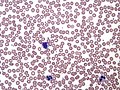
Blood smear is a laboratory procedure that involves spreading a drop of blood thinly onto a glass slide and then staining the blood to view and evaluate the cells. Blood smears are used in the diagnosis and monitoring of many diseases, including anemia, infections, leukemia, lymphoma, and other blood disorders.
Procedure[edit]
The blood smear procedure begins with a clean glass slide. A drop of blood is placed on one end of the slide, and then a spreader slide is used to spread the blood across the slide. This creates a thin layer of blood on the slide, which is then allowed to air dry. Once dry, the slide is stained, typically with a type of stain called a Wright's stain. This stain allows the different types of blood cells to be seen under a microscope.
Interpretation[edit]
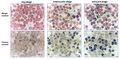
The stained blood smear is examined under a microscope by a pathologist or a laboratory scientist. They look at the size, shape, and number of the different types of blood cells. They also look for any abnormal cells or cell structures. The results of a blood smear can provide important information about a person's health and can help diagnose many different conditions.
Clinical significance[edit]
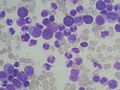
Blood smears are often used in the diagnosis and monitoring of blood disorders and diseases. For example, they can be used to diagnose anemia, in which there are too few red blood cells, or polycythemia, in which there are too many. They can also be used to diagnose infections, as certain types of white blood cells increase in number in response to infection. In addition, blood smears can be used to diagnose and monitor leukemia and lymphoma, as these diseases cause abnormal white blood cells to be present in the blood.
See also[edit]
This medical article is a stub. You can help WikiMD by expanding the page. |
Blood smear[edit]
-
 Blood smear
 -
 Blood smear
 -
 Blood smear
 -
 Blood smear
 -
 Blood smear
 -
 Blood smear
 -
 Blood smear


Medical Disclaimer: WikiMD is for informational purposes only and is not a substitute for professional medical advice. Content may be inaccurate or outdated and should not be used for diagnosis or treatment. Always consult your healthcare provider for medical decisions. Verify information with trusted sources such as CDC.gov and NIH.gov. By using this site, you agree that WikiMD is not liable for any outcomes related to its content. See full disclaimer.
Credits:Most images are courtesy of Wikimedia commons, and templates, categories Wikipedia, licensed under CC BY SA or similar.
Translate page: - East Asian
中文,
日本,
한국어,
South Asian
हिन्दी,
தமிழ்,
తెలుగు,
Urdu,
ಕನ್ನಡ,
Southeast Asian
Indonesian,
Vietnamese,
Thai,
မြန်မာဘာသာ,
বাংলা
European
español,
Deutsch,
français,
Greek,
português do Brasil,
polski,
română,
русский,
Nederlands,
norsk,
svenska,
suomi,
Italian
Middle Eastern & African
عربى,
Turkish,
Persian,
Hebrew,
Afrikaans,
isiZulu,
Kiswahili,
Other
Bulgarian,
Hungarian,
Czech,
Swedish,
മലയാളം,
मराठी,
ਪੰਜਾਬੀ,
ગુજરાતી,
Portuguese,
Ukrainian
